Explore
 Validate
 Learn
Learn Western blot
 Immunohistochemistry
Antibody data
- Antibody Data
- Antigen structure
- References [3]
- Comments [0]
- Validations
- Immunohistochemistry [1]
- Flow cytometry [2]
- Other assay [6]
Submit
Validation data
Reference
Comment
Report error
- Product number
- PA5-26976 - Provider product page

- Provider
- Invitrogen Antibodies
- Product name
- ID4 Polyclonal Antibody
- Antibody type
- Polyclonal
- Antigen
- Synthetic peptide
- Description
- This antibody is predicted to react with porcine based on sequence homology.
- Reactivity
- Human, Mouse
- Host
- Rabbit
- Isotype
- IgG
- Vial size
- 400 μL
- Concentration
- 0.5 mg/mL
- Storage
- Store at 4°C short term. For long term storage, store at -20°C, avoiding freeze/thaw cycles.
Submitted references Pentoxifylline improves the survival of spermatogenic cells via oxidative stress suppression and upregulation of PI3K/AKT pathway in mouse model of testicular torsion-detorsion.
Roles for Kisspeptin in proliferation and differentiation of spermatogonial cells isolated from mice offspring when the cells are cocultured with somatic cells.
Roles for osteocalcin in proliferation and differentiation of spermatogonial cells cocultured with somatic cells.
Dhulqarnain AO, Takzaree N, Hassanzadeh G, Tooli H, Malekzadeh M, Khanmohammadi N, Yaghobinejad M, Solhjoo S, Rastegar T
Heliyon 2021 Apr;7(4):e06868
Heliyon 2021 Apr;7(4):e06868
Roles for Kisspeptin in proliferation and differentiation of spermatogonial cells isolated from mice offspring when the cells are cocultured with somatic cells.
Toolee H, Rastegar T, Solhjoo S, Mortezaee K, Mohammadipour M, Kashani IR, Akbari M
Journal of cellular biochemistry 2019 Apr;120(4):5042-5054
Journal of cellular biochemistry 2019 Apr;120(4):5042-5054
Roles for osteocalcin in proliferation and differentiation of spermatogonial cells cocultured with somatic cells.
Solhjoo S, Akbari M, Toolee H, Mortezaee K, Mohammadipour M, Nematollahi-Mahani SN, Shahrokhi A, Sayadi M, Rastegar T
Journal of cellular biochemistry 2019 Apr;120(4):4924-4934
Journal of cellular biochemistry 2019 Apr;120(4):4924-4934
No comments: Submit comment
Supportive validation
- Submitted by
- Invitrogen Antibodies (provider)
- Main image
- Experimental details
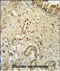
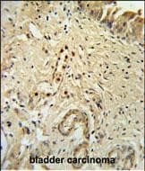
- Immunohistochemistry analysis of ID4 in formalin fixed and paraffin embedded bladder carcinoma. Samples were incubated with ID4 polyclonal antibody (Product # PA5-26976) followed by peroxidase conjugation of the secondary antibody and DAB staining. This data demonstrates the use of this antibody for immunohistochemistry. Clinical relevance has not been evaluated.
Supportive validation
- Submitted by
- Invitrogen Antibodies (provider)
- Main image

- Experimental details
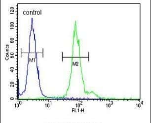
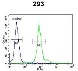
- Flow cytometry analysis of 293 cells using an ID4 polyclonal antibody (Product # PA5-26976) (right) compared to a negative control cell (left) at a dilution of 1:10-50, followed by a FITC-conjugated goat anti-rabbit antibody
- Submitted by
- Invitrogen Antibodies (provider)
- Main image
- Experimental details
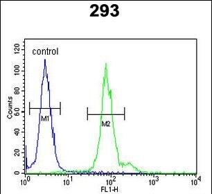
- Flow cytometry of ID4 in 293 cells (right histogram). Samples were incubated with ID4 polyclonal antibody (Product # PA5-26976) followed by FITC-conjugated goat-anti-rabbit secondary antibody. Negative control (left histogram).
Supportive validation
- Submitted by
- Invitrogen Antibodies (provider)
- Main image
- Experimental details
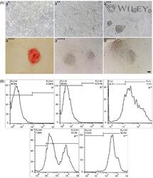
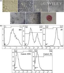
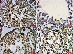
- 1 A, Morphological and identity of spermatogonial cells (SCs). Large and dense colonies are visible in an image taken by an inverted microscope at the end of the first week of culture (a'''). Results from alkaline phosphatase staining indicate apparent cellular colonies after culturing for 14 (a'''') and 28 (a'''''') days (Scale bar = 100 mum). a', day 1; a'', day 4; and; a''''', day 21. B, ID4, alpha6-, and beta1-integrin were assessed by the flow cytometry to determine whether the isolated cells had SC identity. As it is evident, these markers were expressed at high percentages in the cells. b' and b'', control; b''', ID4; b'''', alpha6-integrin; and b''''', beta1-integrin
- Submitted by
- Invitrogen Antibodies (provider)
- Main image
- Experimental details
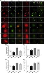
- 3 A: Immunocytochemistry (ICC) of MVH and ID4 as proliferation-related markers in spermatogonial cells (SCs) cocultured with Leydig cell to evaluate to role for osteocalcin. The markers were stained green, and nuclei stained red in the cells (Scale bars = 100 mum). B,C: Quantification of the ICC data showed no significant expressions for MVH and ID4 in the osteocalcin-treated cells. Control, a-a'''', c-c'''', e-e''', and g-g'''; and osteocalcin, b-b'''', d-d'''', f-f''', and h-h'''

- Submitted by
- Invitrogen Antibodies (provider)
- Main image
- Experimental details
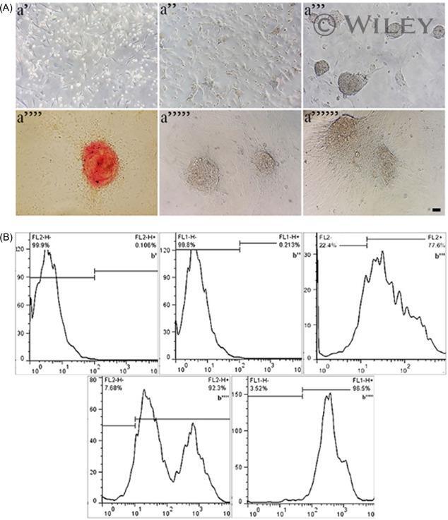
- 1 Spermatogonial cell (SC) morphology and identity. SCs growing in the culture showed large and compact colonies by the end of the first week. a', day 1; a'', day 4; a''', day 7; a'''', day 14; a''''', day 21; and a'''''', day 28. a''''''' shows red staining colonies of SCs after alkaline phosphatase staining (scale bars = 100 mum). B, flow cytometry analysis for SC markers ID4 (b'), alpha6 integrin (b'') and beta1 integrin (b'''). Negative controls for FITC and PE were marker by respective b'''' and b'''''. PE, phycoerythrin

- Submitted by
- Invitrogen Antibodies (provider)
- Main image
- Experimental details
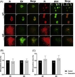
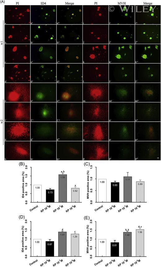
- 3 Immunofluorescence staining of spermatogonial cells (SCs) morphology for identifying proliferative (MVH and ID4) markers assessed at the end of weeks 1 and 2. Green stained SCs shows expression of the markers in the cells. Nuclei were stained red in the cells (Scale bars = 50 mum). Graphs are indicative of the quantified area stained fluorescently for the markers. a-a'''' e-e'''' i-i'''' m-m'''' control; b-b'''' f-f'''' j-j'''' n-n'''' Kp 10 -8 M; c-c'''' g-g'''' k-k'''' o-o'''' Kp 10 -7 M; and d-d'''' h-h'''' l-l'''' p-p'''' Kp 10 -6 M. a = P
- Submitted by
- Invitrogen Antibodies (provider)
- Main image
- Experimental details
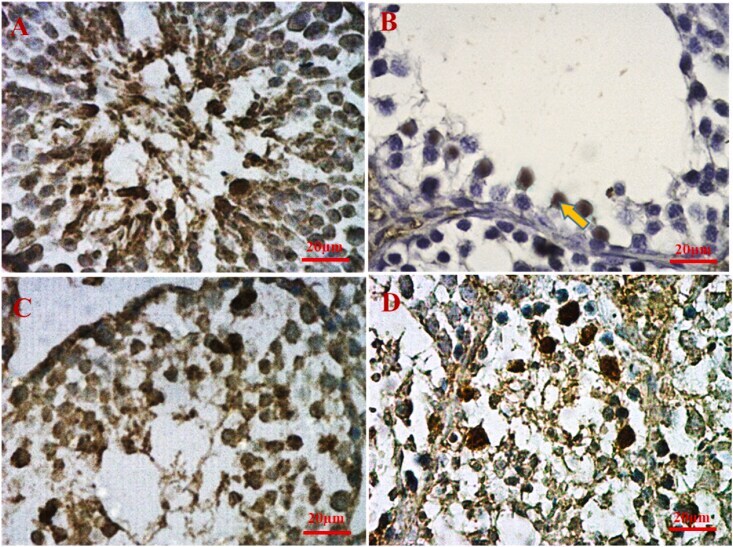
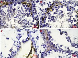
- Figure 2 Immunohistochemistry staining for ID4 protein: A: Control with normal expression of ID4 protein in the seminiferous tubule, B: T1 (torsion-detorsion + s ingle dose of 100 mg/kg PTX with significant expression of ID4 protein (yellow arrow)), C: T2 (torsion-detorsion + 25 mg/kg/day PTX for two weeks with low expression of ID4 protein, D: T/D (torsion-detorsion without any treatment and with significantly low expression of ID4 protein. (H & E, scale bar: 20 mum). (Full, non-unadjusted images are shown as supplementary material). Figure 2
- Submitted by
- Invitrogen Antibodies (provider)
- Main image
- Experimental details
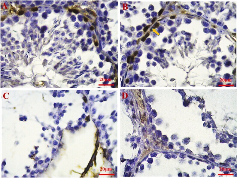
- Figure 3 Immunohistochemistry staining for SCP3 protein; A: Control (with normal expression of ID4 protein in the seminiferous tubule), B: T1 (torsion-detorsion + single dose 100 mg/kg PTX with significant increase in the expression of scp3 protein (Yellow arrow)), C: T2 (torsion-detorsion + 25 mg/kg/day PTX for two weeks with significant increase in expression of sp3 protein, D: T/D (torsion-detorsion without any treatment and with significantly low expression of scp3 protein. (H & E, scale bar: 20 mum). (Full, non-unadjusted images are shown as supplementary material). Figure 3