 Explore
 Validate
 Learn
LearnPA5-47036
antibody from Invitrogen Antibodies
Targeting: BIRC2
API1, c-IAP1, cIAP1, hiap-2, MIHB, RNF48
 Western blot
Antibody data
- Antibody Data
- Antigen structure
- References [0]
- Comments [0]
- Validations
- Western blot [6]
- Immunocytochemistry [1]
- Immunohistochemistry [2]
Submit
Validation data
Reference
Comment
Report error
- Product number
- PA5-47036 - Provider product page

- Provider
- Invitrogen Antibodies
- Product name
- cIAP1 Polyclonal Antibody
- Antibody type
- Polyclonal
- Antigen
- Recombinant full-length protein
- Description
- This antibody does not cross-react with recombinant human cIAP-2 or XIAP.
- Concentration
- 0.2 mg/mL
No comments: Submit comment
Supportive validation
- Submitted by
- Invitrogen Antibodies (provider)
- Main image

- Experimental details
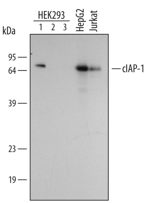
- Western blot analysis from lysates of HEK293 human embryonic kidney cell line transfected with human cIAP-1 (lane 1), human cIAP-2 (lane 2), or non-transfected (lane 3). PVDF membrane was probed with 0.5 µg/mL Goat Anti-human cIAP-1/HIAP-2 Antigen Affinity-purified Polyclonal Antibody (Product # PA5-47036) followed by HRP-conjugated Anti-Goat IgG Secondary Antibody. For additional reference, lystates of HepG2 human hepatocellular carcinoma cell line and Jurkat human acute T cell leukemia cell line were included. A specific band for cIAP-1/HIAP-2 was detected at approximately 65 kDa (as indicated). This experiment was conducted under reducing conditions.
- Submitted by
- Invitrogen Antibodies (provider)
- Main image
- Experimental details
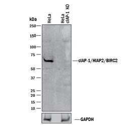
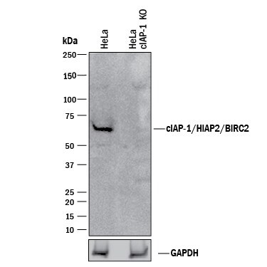
- Knockout validation by Western blot analysis of cIAP1 in lysates of HeLa human cervical epithelial carcinoma parental cell line and cIAP-1/HIAP-2 knockout HeLa cell line (KO). Samples were incubated in cIAP1 polyclonal antibody (Product # PA5-47036) using a dilution of 0.5 µg/mL followed by a HRP-conjugated Anti-Goat IgG secondary antibody. A specific band was detected for cIAP‚1/HIAP‚2 at approximately 68 kDa (as indicated) in the parental HeLa cell line, but is not detectable in knockout HeLa cell line. GAPDH is shown as a loading control. This experiment was conducted under reducing conditions.
- Submitted by
- Invitrogen Antibodies (provider)
- Main image

- Experimental details
- Western blot analysis of cIAP1 in HEK293 human embryonic kidney cell line transfected with human cIAP-1 (lane 1), human cIAP-2 (lane 2), or non-transfected (lane 3). Samples were incubated in cIAP1 polyclonal antibody (Product # PA5-47036) using a dilution of 0.5 µg/mL followed by a HRP-conjugated Anti-Goat IgG secondary antibody. For additional reference, lystates of HepG2 human hepatocellular carcinoma cell line and Jurkat human acute T cell leukemia cell line were included. A specific band for cIAP‚1/HIAP‚2 was detected at approximately 65 kDa (as indicated). This experiment was conducted under reducing conditions.

- Submitted by
- Invitrogen Antibodies (provider)
- Main image
- Experimental details
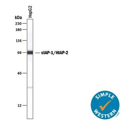
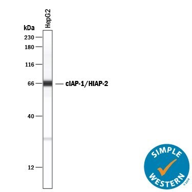
- Western blot analysis of cIAP1 in 0.5 mg/mL lysates of HepG2 human hepatocellular carcinoma cell line. Samples were incubated in cIAP1 polyclonal antibody (Product # PA5-47036) using a dilution of 5 µg/mL followed by HRP-conjugated Anti-Goat IgG at a dilution of 0.0763888888888889. A specific band was detected for cIAP‚1/HIAP‚2 at approximately 66 kDa (as indicated). This experiment was conducted under reducing conditions and using the 12-230 kDa separation system.
- Submitted by
- Invitrogen Antibodies (provider)
- Main image

- Experimental details
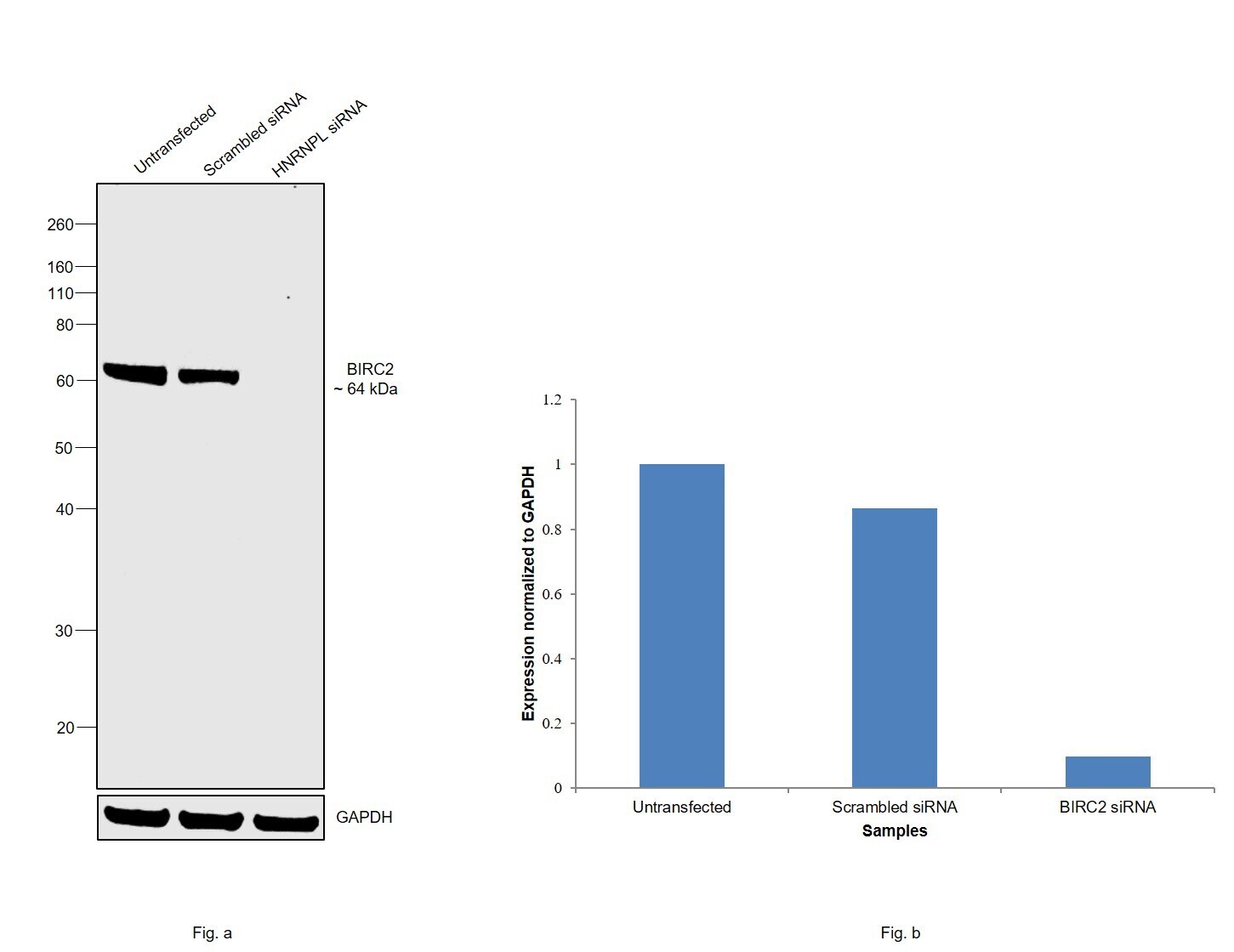
- Knockdown of BIRC2 was achieved by transfecting HeLa with BIRC2 specific siRNAs (Silencer® select Product # s1450). Western blot analysis (Fig. a) was performed using modified whole cell extracts (1% SDS) from the BIRC2 knockdown cells (lane 3), non-specific scrambled siRNA transfected cells (lane 2) and untransfected cells (lane 1). The blot was probed with cIAP1 Polyclonal Antibody (Product # PA5-47036, 0.5 µg/ml) and Goat anti-Rabbit IgG (H+L) Superclonal™ Recombinant Secondary Antibody, HRP (Product # A27036, 1:4000 dilution). Densitometric analysis of this western blot is shown in histogram (Fig. b). Decrease in signal upon siRNA mediated knock down confirms that antibody is specific to BIRC2.
- Submitted by
- Invitrogen Antibodies (provider)
- Main image

- Experimental details
- Western blot was performed using Anti-cIAP1 Polyclonal Antibody (Product # PA5-47036) and a 64 kDa corresponding to BIRC2 was observed across cell lines tested. Modified whole cell extracts (1% SDS) (30 µg lysate) of HeLa (Lane 1), Hep G2 (Lane 2), HCT116 (Lane 3), T-47D (Lane 4), MCF-7 (Lane 5), MDA-MB-231 (Lane 6), A549 (Lane 7), LNCaP (Lane 8) and Jurkat (Lane 9) were electrophoresed using Novex® NuPAGE® 4-12 % Bis-Tris gel (Product # NP0322BOX). Resolved proteins were then transferred onto a nitrocellulose membrane (Product # IB23001) by iBlot® 2 Dry Blotting System (Product # IB21001). The blot was probed with the primary antibody (0.5 µg/ml) and detected by chemiluminescence with Rabbit anti-Goat IgG (H+L) Superclonal™ Recombinant Secondary Antibody, HRP (Product # A27014, 1:4000 dilution) using the iBright FL 1000 (Product # A32752). Chemiluminescent detection was performed using Novex® ECL Chemiluminescent Substrate Reagent Kit (Product # WP20005)..

Supportive validation
- Submitted by
- Invitrogen Antibodies (provider)
- Main image
- Experimental details
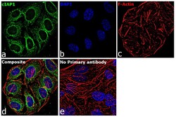
- Immunofluorescence analysis of cIAP1 was performed using 70% confluent log phase HeLa cells. The cells were fixed with 4% paraformaldehyde for 10 minutes, permeabilized with 0.1% Triton™ X-100 for 15 minutes, and blocked with 2% BSA for 1 hour at room temperature. The cells were labeled with cIAP1 Goat Polyclonal Antibody (Product # PA5-47036) at 5 µg/mL in 0.1% BSA, incubated at 4 degree celsius overnight and then labeled with Rabbit anti-Goat IgG (H+L) Cross-Adsorbed Secondary Antibody, Alexa Fluor 488 (Product # A-11078) at a dilution of 1:2000 for 45 minutes at room temperature (Panel a: green). Nuclei (Panel b: blue) were stained with ProLong™ Diamond Antifade Mountant with DAPI (Product # P36962). F-actin (Panel c: red) was stained with Rhodamine Phalloidin (Product # R415). Panel d represents the merged image showing Cytoplasmic localization. Panel e represents control cells with no primary antibody to assess background. The images were captured at 60X magnification.

Supportive validation
- Submitted by
- Invitrogen Antibodies (provider)
- Main image

- Experimental details
- Immunohistochemical analysis of cIAP1 in immersion fixed paraffin-embedded sections of human lymphoma. Samples were incubated in cIAP1 polyclonal antibody (Product # PA5-47036) using a dilution of 15 µg/mL overnight at 4 °C. Tissue was stained using the Anti-Goat HRP-DAB Cell & Tissue Staining Kit (brown) and counterstained with hematoxylin (blue). Lower panel shows a lack of labeling if primary antibodies are omitted and tissue is stained only with secondary antibody followed by incubation with detection reagents.

- Submitted by
- Invitrogen Antibodies (provider)
- Main image

- Experimental details
- Immunohistochemical analysis of cIAP1 in immersion fixed paraffin-embedded sections of human lymph node. Samples were incubated in cIAP1 polyclonal antibody (Product # PA5-47036) using a dilution of 10 µg/mL overnight at 4 °C. Tissue was stained using the Anti-Goat HRP-DAB Cell & Tissue Staining Kit (brown) and counterstained with hematoxylin (blue). Specific staining was localized to lymphocytes.
