Explore
 Validate
 Learn
LearnA01700-1
antibody from Boster Biological Technology
Targeting: BIRC2
API1, c-IAP1, cIAP1, hiap-2, MIHB, RNF48
 Western blot
 ELISA
ELISAAntibody data
- Antibody Data
- Antigen structure
- References [0]
- Comments [0]
- Validations
- Western blot [1]
Submit
Validation data
Reference
Comment
Report error
- Product number
- A01700-1 - Provider product page

- Provider
- Boster Biological Technology
- Product name
- Anti-cIAP1/BIRC2 Antibody Picoband™
- Antibody type
- Polyclonal
- Description
- Polyclonal antibody for CIAP1/BIRC2 detection. Host: Rabbit.Size: 100μg/vial. Tested applications: ELISA. Reactive species: Human. CIAP1/BIRC2 information: Subcellular Localization: Cytoplasm. Nucleus. Agents that induce either the extrinsic or intrinsic apoptotic pathways promote its redistribution from the nuclear compartment to the cytoplasmic compartment. Associated with the midbody in telophase cells, and found diffusely in the nucleus of interphase cells; Tissue Specificity: Present in many fetal and adult tissues. Mainly expressed in adult skeletal muscle, thymus, testis, ovary, and pancreas, low or absent in brain and peripheral blood leukocytes.
- Reactivity
- Human, Mouse, Rat
- Host
- Rabbit
- Vial size
- 100μg/vial
- Concentration
- 0.5-1mg/ml, actual concentration vary by lot. Use suggested dilution ratio to decide dilution procedure.
- Storage
- At -20°C for one year. After reconstitution, at 4°C for one month. It can also be aliquoted and stored frozen at -20°C for a longer time. Avoid repeated freezing and thawing.
- Handling
- Add 0.2ml of distilled water will yield a concentration of 500ug/ml.
No comments: Submit comment
Supportive validation
- Submitted by
- Boster Biological Technology (provider)
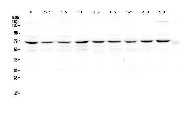
- Main image
- Experimental details
- Western blot analysis of cIAP1 using anti-cIAP1 antibody (A01700-1). Electrophoresis was performed on a 5-20% SDS-PAGE gel at 70V (Stacking gel) / 90V (Resolving gel) for 2-3 hours. The sample well of each lane was loaded with 50ug of sample under reducing conditions. Lane 1: human Hela whole cell lysates,Lane 2: human Jurkat whole cell lysates,Lane 3: human MCF-7 whole cell lysates,Lane 4: human COLO-320 whole cell lysates,Lane 5: human U-87MG whole cell lysates,Lane 6: human A549 whole cell lysates,Lane 7: rat thymus tissue lysates,Lane 8: mouse thymus tissue lysates,Lane 9: mouse testis tissue lysates. After Electrophoresis, proteins were transferred to a Nitrocellulose membrane at 150mA for 50-90 minutes. Blocked the membrane with 5% Non-fat Milk/ TBS for 1.5 hour at RT. The membrane was incubated with rabbit anti-cIAP1 antigen affinity purified polyclonal antibody (Catalog # A01700-1) at 0.5 μg/mL overnight at 4°C, then washed with TBS-0.1%Tween 3 times with 5 minutes each and probed with a goat anti-rabbit IgG-HRP secondary antibody at a dilution of 1:10000 for 1.5 hour at RT. The signal is developed using an Enhanced Chemiluminescent detection (ECL) kit (Catalog # EK1002) with Tanon 5200 system. A specific band was detected for cIAP1 at approximately 70KD. The expected band size for cIAP1 is at 70KD.
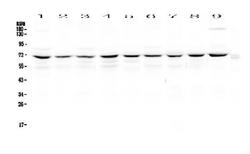
- Additional image