Explore
 Validate
 Learn
Learn Western blot
Antibody data
- Antibody Data
- Antigen structure
- References [3]
- Comments [0]
- Validations
- Western blot [1]
- Immunohistochemistry [1]
Submit
Validation data
Reference
Comment
Report error
- Product number
- sc-12739 - Provider product page

- Provider
- Santa Cruz Biotechnology
- Proper citation
- Santa Cruz Biotechnology Cat#sc-12739, RRID:AB_626792
- Product name
- Anti-CSNK2B
- Antibody type
- Monoclonal
- Reactivity
- Human
- Host
- Mouse
Submitted references Three novel downstream promoter elements regulate MHC class I promoter activity in mammalian cells.
Arsenic-induced malignant transformation of human keratinocytes: involvement of Nrf2.
Mechanistic studies of the mitotic activation of Mos.
Lee N, Iyer SS, Mu J, Weissman JD, Ohali A, Howcroft TK, Lewis BA, Singer DS
PloS one 2010 Dec 13;5(12):e15278
PloS one 2010 Dec 13;5(12):e15278
Arsenic-induced malignant transformation of human keratinocytes: involvement of Nrf2.
Pi J, Diwan BA, Sun Y, Liu J, Qu W, He Y, Styblo M, Waalkes MP
Free radical biology & medicine 2008 Sep 1;45(5):651-8
Free radical biology & medicine 2008 Sep 1;45(5):651-8
Mechanistic studies of the mitotic activation of Mos.
Yue J, Ferrell JE Jr
Molecular and cellular biology 2006 Jul;26(14):5300-9
Molecular and cellular biology 2006 Jul;26(14):5300-9
No comments: Submit comment
Supportive validation
- Submitted by
- per
- Main image

- Experimental details
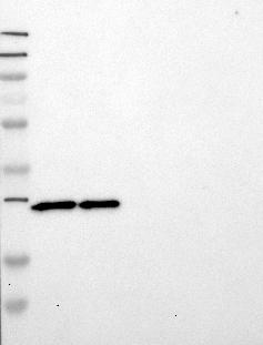
- Western blot analysis of antibody specificity using a routine panel composed of IgG/HSA-depleted human plasma and protein lysates from selected human tissues and cell lines.
- Validation comment
- Single band corresponding to the predicted size in kDa (+/-20%).
- Primary Ab dilution
- 1:500
- Secondary Ab dilution
- 1:7000
- Lane 1
- Marker [kDa]: 250, 130, 95, 72, 55, 36, 28, 17, 11
- Lane 2
- RT-4
- Lane 3
- U-251MG sp
- Lane 4
- Human Plasma
- Lane 5
- Liver
- Lane 6
- Tonsil
- Theoretical target weight
- [kDa] 27
Supportive validation
- Submitted by
- per
- Main image
- Experimental details
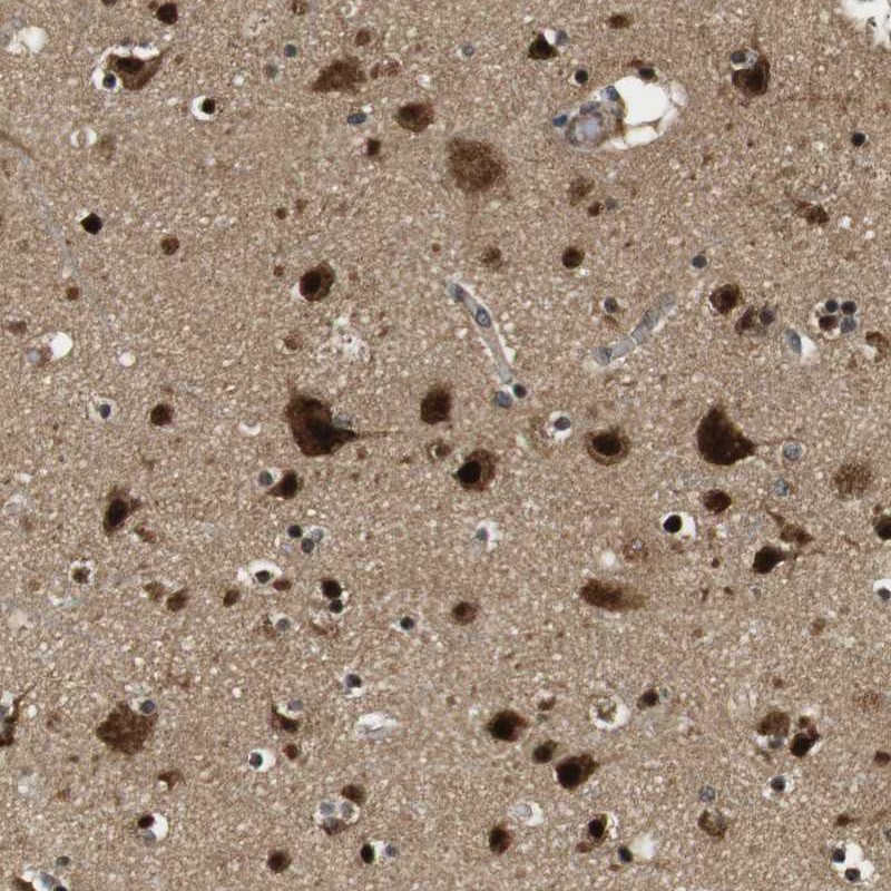
- Immunohistochemical staining of human cerebral cortex shows strong cytoplasmic as well as nuclear positivity in neuronal cells and glial cells.
- Validation comment
- Staining pattern consistent with experimental and/or bioinformatic data.