Explore
 Validate
 Learn
Learn Western blot
Antibody data
- Antibody Data
- Antigen structure
- References [2]
- Comments [0]
- Validations
- Western blot [2]
- Immunocytochemistry [4]
- Immunohistochemistry [3]
- Chromatin Immunoprecipitation [2]
- Other assay [2]
Submit
Validation data
Reference
Comment
Report error
- Product number
- PA5-27416 - Provider product page

- Provider
- Invitrogen Antibodies
- Product name
- CK2 beta Polyclonal Antibody
- Antibody type
- Polyclonal
- Antigen
- Recombinant full-length protein
- Description
- Recommended positive controls: MCF-7, NIH-3T3. Predicted reactivity: Mouse (100%), Rat (100%), Zebrafish (100%), Xenopus laevis (100%), Pig (100%), Rabbit (100%), Chicken (100%), Sheep (100%), Rhesus Monkey (100%), Bovine (100%). Store product as a concentrated solution. Centrifuge briefly prior to opening the vial.
- Reactivity
- Human, Mouse
- Host
- Rabbit
- Isotype
- IgG
- Vial size
- 100 μL
- Concentration
- 0.15 mg/mL
- Storage
- Store at 4°C short term. For long term storage, store at -20°C, avoiding freeze/thaw cycles.
Submitted references Depletion of Numb and Numblike in Murine Lung Epithelial Cells Ameliorates Bleomycin-Induced Lung Fibrosis by Inhibiting the β-Catenin Signaling Pathway.
A Herpesviral induction of RAE-1 NKG2D ligand expression occurs through release of HDAC mediated repression.
Ianni A, Hofmann M, Kumari P, Tarighi S, Al-Tamari HM, Görgens A, Giebel B, Nolte H, Krüger M, Salwig I, Pullamsetti SS, Günther A, Schneider A, Braun T
Frontiers in cell and developmental biology 2021;9:639162
Frontiers in cell and developmental biology 2021;9:639162
A Herpesviral induction of RAE-1 NKG2D ligand expression occurs through release of HDAC mediated repression.
Greene TT, Tokuyama M, Knudsen GM, Kunz M, Lin J, Greninger AL, DeFilippis VR, DeRisi JL, Raulet DH, Coscoy L
eLife 2016 Nov 22;5
eLife 2016 Nov 22;5
No comments: Submit comment
Supportive validation
- Submitted by
- Invitrogen Antibodies (provider)
- Main image

- Experimental details
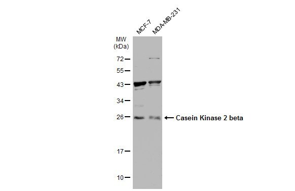
- Western Blot analysis of CK2 beta was performed by separating 30 µg of various whole cell extracts by 12% SDS-PAGE. Proteins were transferred to a membrane and probed with a CK2 beta Polyclonal Antibody (Product # PA5-27416) at a dilution of 1:500 and a HRP-conjugated anti-rabbit IgG secondary antibody.
- Submitted by
- Invitrogen Antibodies (provider)
- Main image
- Experimental details
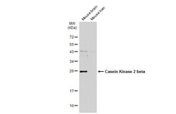
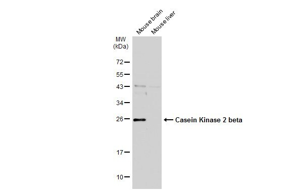
- Western blot analysis of CK2 beta was performed by separating 50 µg of various tissue extracts by 12% SDS-PAGE. Proteins were transferred to a membrane and probed with a CK2 beta Polyclonal Antibody (Product # PA5-27416) at a dilution of 1:500. The HRP-conjugated anti-rabbit IgG antibody was used to detect the primary antibody.
Supportive validation
- Submitted by
- Invitrogen Antibodies (provider)
- Main image
- Experimental details
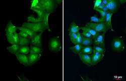
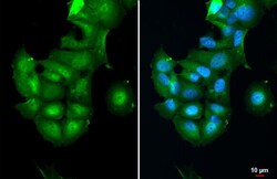
- Immunofluorescent analysis of Casein Kinase 2 beta in paraformaldehyde-fixed HeLa cells using a Casein Kinase 2 beta polyclonal antibody (Product # PA5-27416) at a 1:500 dilution.
- Submitted by
- Invitrogen Antibodies (provider)
- Main image
- Experimental details
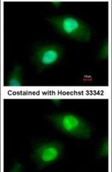
- CK2 beta Polyclonal Antibody detects Casein Kinase 2 beta protein at cytoplasm and nucleus by immunofluorescent analysis. Sample: MCF-7 cells were fixed in 4% paraformaldehyde at RT for 15 min. Green: Casein Kinase 2 beta stained by CK2 beta Polyclonal Antibody (Product # PA5-27416) diluted at 1:500.

- Submitted by
- Invitrogen Antibodies (provider)
- Main image

- Experimental details
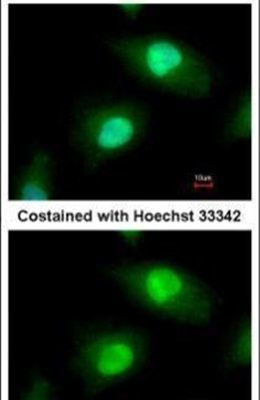
- CK2 beta Polyclonal Antibody detects Casein Kinase 2 beta protein at cytoplasm and nucleus by immunofluorescent analysis. Sample: MCF-7 cells were fixed in 4% paraformaldehyde at RT for 15 min. Green: Casein Kinase 2 beta stained by CK2 beta Polyclonal Antibody (Product # PA5-27416) diluted at 1:500.

- Submitted by
- Invitrogen Antibodies (provider)
- Main image
- Experimental details
- CK2 beta Polyclonal Antibody detects Casein Kinase 2 beta protein at cytoplasm and nucleus by immunofluorescent analysis. Sample: MCF-7 cells were fixed in 4% paraformaldehyde at RT for 15 min. Green: Casein Kinase 2 beta stained by CK2 beta Polyclonal Antibody (Product # PA5-27416) diluted at 1:500.

Supportive validation
- Submitted by
- Invitrogen Antibodies (provider)
- Main image

- Experimental details
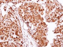
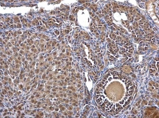
- CK2 beta Polyclonal Antibody detects Casein Kinase 2 beta protein at nucleus and cytosol on mouse ovary by immunohistochemical analysis. Sample: Paraffin-embedded mouse ovary. CK2 beta Polyclonal Antibody (Product # PA5-27416) dilution: 1:500. Antigen Retrieval: EDTA based buffer, pH 8.0, 15 min.

- Submitted by
- Invitrogen Antibodies (provider)
- Main image
- Experimental details
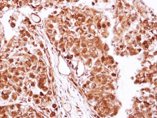
- Immunohistochemical analysis of paraffin-embedded CL1-0 xenograft, using Casein Kinase 2 beta (Product # PA5-27416) antibody at 1:100 dilution. Antigen Retrieval: EDTA based buffer, pH 8.0, 15 min.
- Submitted by
- Invitrogen Antibodies (provider)
- Main image

- Experimental details
- CK2 beta Polyclonal Antibody detects Casein Kinase 2 beta protein at nucleus and cytosol on mouse ovary by immunohistochemical analysis. Sample: Paraffin-embedded mouse ovary. CK2 beta Polyclonal Antibody (Product # PA5-27416) dilution: 1:500. Antigen Retrieval: EDTA based buffer, pH 8.0, 15 min.
Supportive validation
- Submitted by
- Invitrogen Antibodies (provider)
- Main image
- Experimental details
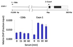
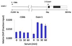
- Chromatin immunoprecipitation analysis of Casein Kinase II beta was performed using cross-linked chromatin from 1 x 10⁶ HCT116 colon carcinoma cells treated with serum for 0, 15, 30, and 60 minutes. Immunoprecipitation was performed using a multiplex microplate Matrix ChIP assay (see reference for Matrix ChIP protocol: http://www.ncbi.nlm.nih.gov/pubmed/22098709) with 1.0 µL/100 µL well volume of a Casein Kinase II beta polyclonal antibody (Product # PA5-27416). Chromatin aliquots from ~1 x 10⁵ cells were used per ChIP pull-down. Quantitative PCR data were done in quadruplicate using 1 µL of eluted DNA in 2 µL SYBR real-time PCR reactions containing primers to amplify -15kb upstream of the Egr1 gene or exon-1 of Egr1. PCR calibration curves were generated for each primer pair from a dilution series of sheared total genomic DNA. Quantitation of immunoprecipitated chromatin is presented as signal relative to the total amount of input chromatin. Results represent the mean +/- SEM for three experiments. A schematic representation of the Egr-1 locus is shown above the data where boxes represent exons (black boxes = translated regions, white boxes = untranslated regions); the zigzag line represents an intron; and the straight line represents upstream sequence. Regions amplified by Egr-1 primers are represented by black bars. Data courtesy of the Innovators Program.

- Submitted by
- Invitrogen Antibodies (provider)
- Main image
- Experimental details
- Chromatin immunoprecipitation analysis of Casein Kinase II beta was performed using cross-linked chromatin from 1 x 10⁶ HCT116 colon carcinoma cells treated with serum for 0, 15, 30, and 60 minutes. Immunoprecipitation was performed using a multiplex microplate Matrix ChIP assay (see reference for Matrix ChIP protocol: http://www.ncbi.nlm.nih.gov/pubmed/22098709) with 1.0 µL/100 µL well volume of a Casein Kinase II beta polyclonal antibody (Product # PA5-27416). Chromatin aliquots from ~1 x 10⁵ cells were used per ChIP pull-down. Quantitative PCR data were done in quadruplicate using 1 µL of eluted DNA in 2 µL SYBR real-time PCR reactions containing primers to amplify -15kb upstream of the Egr1 gene or exon-1 of Egr1. PCR calibration curves were generated for each primer pair from a dilution series of sheared total genomic DNA. Quantitation of immunoprecipitated chromatin is presented as signal relative to the total amount of input chromatin. Results represent the mean +/- SEM for three experiments. A schematic representation of the Egr-1 locus is shown above the data where boxes represent exons (black boxes = translated regions, white boxes = untranslated regions); the zigzag line represents an intron; and the straight line represents upstream sequence. Regions amplified by Egr-1 primers are represented by black bars. Data courtesy of the Innovators Program.

Supportive validation
- Submitted by
- Invitrogen Antibodies (provider)
- Main image

- Experimental details
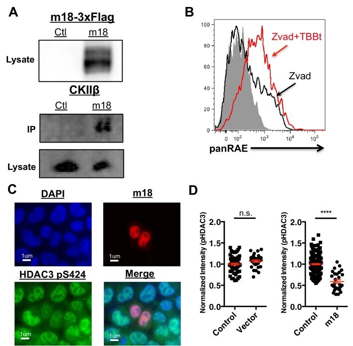
- Figure 6. CK2 directly interacts with m18 and represses HDAC function. ( A ) Immunoprecipitation (IP) of m18 was performed in lysates of cells expressing m18-3xFlag or empty 3xFlag vector, and the products were analyzed for FLAG and CK2beta by western blot. Data are representative of three independent experiments. ( B ) Mouse fibroblasts were treated with CK2 inhibitor TBBt in conjunction with zVAD or zVAD alone and analyzed for RAE-1 expression by flow cytometry. Data is representative of three independent experiments. ( C ) Representative image of fibroblasts expressing m18-RFP and stained for HDAC3 pS424. ( D ) Quantification of HDAC3 pS424 levels in cells expressing m18-RFP or RFP control vector from compared to non-transfected controls in same field of view. Red bars are representative of mean+-SEM. Data are representative of three independent experiments. ****p
- Submitted by
- Invitrogen Antibodies (provider)
- Main image
- Experimental details
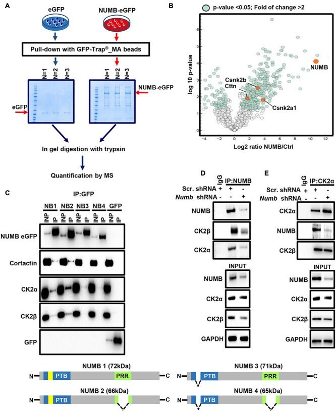
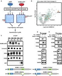
- FIGURE 4 NUMB interacts with different proteins including CK2. (A) A scheme depicting the experimental approach used for mass spectrometry-based analysis of NUMB-eGFP interactor partners. (B) A volcano plot showing NUMB-interacting proteins detected by mass spectrometry. (C) Coupled immunoprecipitation (GFP antibody) and western blot analysis (GFP, cortactin, CK2alpha, and CK2beta antibodies) of stable NUMB 1-4-overexpressing cells ( n = 3; upper panel). The lower scheme illustrates the structure of the four NUMB isoforms (PTB, phospho-tyrosine-binding domain; PRR, proline-rich domain; yellow box, amino acid insert in the PTB domain) that might be responsible for interaction with specific targets. See text for details. (D) Coupled immunoprecipitation (NUMB antibody) and western blot analysis (NUMB, CK2alpha, and CK2beta antibodies) of scramble and Numb KD cells. Non-immune immunoglobulin (IgG) was used as a negative control ( n = 3). (E) Coupled immunoprecipitation (CK2alpha antibody) and western blot analysis (NUMB and CK2beta antibodies) of scramble and Numb KD cells.