Explore
 Validate
 Learn
Learn Immunohistochemistry
Antibody data
- Antibody Data
- Antigen structure
- References [3]
- Comments [0]
- Validations
- Immunohistochemistry [1]
Submit
Validation data
Reference
Comment
Report error
- Product number
- HPA002112 - Provider product page

- Provider
- Atlas Antibodies
- Proper citation
- Atlas Antibodies Cat#HPA002112, RRID:AB_1857412
- Product name
- Anti-HYAL1
- Antibody type
- Polyclonal
- Description
- Polyclonal Antibody against Human HYAL1, Gene description: hyaluronoglucosaminidase 1, Alternative Gene Names: FUS2, HYAL-1, LUCA1, NAT6, Validated applications: IHC, Uniprot ID: Q12794, Storage: Store at +4°C for short term storage. Long time storage is recommended at -20°C.
- Reactivity
- Human
- Host
- Rabbit
- Conjugate
- Unconjugated
- Isotype
- IgG
- Vial size
- 100 µl
- Concentration
- 0.2 mg/ml
- Storage
- Store at +4°C for short term storage. Long time storage is recommended at -20°C.
- Handling
- The antibody solution should be gently mixed before use.
Submitted references The Hyaluronic Acid System is Intact in Menstrual Endometrial Cells in Women With and Without Endometriosis
Evaluation of protein biomarkers of prostate cancer aggressiveness
Inverse expression of hyaluronidase 2 and hyaluronan synthases 1-3 is associated with reduced hyaluronan content in malignant cutaneous melanoma.
Knudtson J, McLaughlin J, Santos M, Binkley P, Tekmal R, Schenken R
Reproductive Sciences 2018;26(1):109-113
Reproductive Sciences 2018;26(1):109-113
Evaluation of protein biomarkers of prostate cancer aggressiveness
Rizzardi A, Rosener N, Koopmeiners J, Isaksson Vogel R, Metzger G, Forster C, Marston L, Tiffany J, McCarthy J, Turley E, Warlick C, Henriksen J, Schmechel S
BMC Cancer 2014;14(1)
BMC Cancer 2014;14(1)
Inverse expression of hyaluronidase 2 and hyaluronan synthases 1-3 is associated with reduced hyaluronan content in malignant cutaneous melanoma.
Siiskonen H, Poukka M, Tyynelä-Korhonen K, Sironen R, Pasonen-Seppänen S
BMC cancer 2013 Apr 5;13:181
BMC cancer 2013 Apr 5;13:181
No comments: Submit comment
Supportive validation
- Submitted by
- Atlas Antibodies (provider)
- Enhanced method
- Orthogonal validation
- Main image
- Experimental details
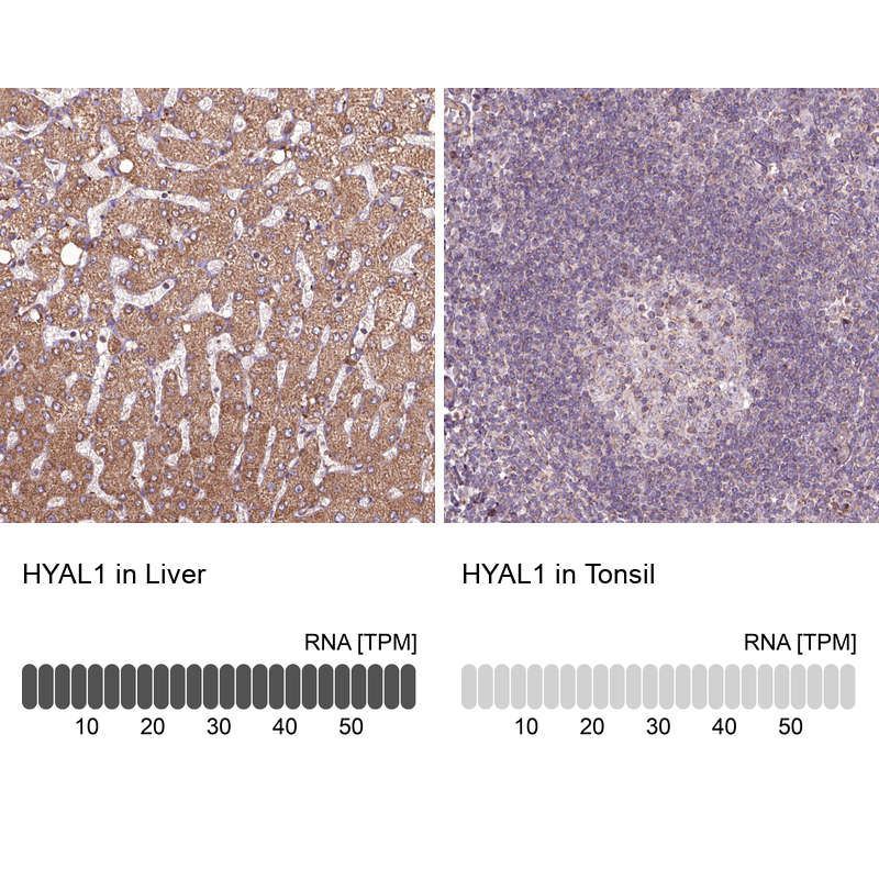
- Immunohistochemistry analysis in human liver and tonsil tissues using HPA002112 antibody. Corresponding HYAL1 RNA-seq data are presented for the same tissues.
- Sample type
- Human
- Protocol
- Protocol