Explore
 Validate
 Learn
Learn Western blot
Antibody data
- Antibody Data
- Antigen structure
- References [0]
- Comments [0]
- Validations
- Western blot [3]
- Immunohistochemistry [1]
- Flow cytometry [2]
Submit
Validation data
Reference
Comment
Report error
- Product number
- MA5-18292 - Provider product page

- Provider
- Invitrogen Antibodies
- Product name
- ARID5A Monoclonal Antibody (P18112)
- Antibody type
- Monoclonal
- Antigen
- Recombinant full-length protein
- Description
- Recommended positive controls: 3xFlag-human ARID5A-transfected 293T, THP-1, THP-1 nuclear extract, Raw264.7, Raw264.7 (50 ng/mL LPS, 4hr). Predicted reactivity: Mouse (83%), Bovine (88%). Store product as a concentrated solution. Centrifuge briefly prior to opening the vial.
- Reactivity
- Human, Mouse
- Host
- Mouse
- Isotype
- IgG
- Antibody clone number
- P18112
- Vial size
- 100 μL
- Concentration
- 1 mg/mL
- Storage
- Store at 4°C short term. For long term storage, store at -20°C, avoiding freeze/thaw cycles.
No comments: Submit comment
Supportive validation
- Submitted by
- Invitrogen Antibodies (provider)
- Main image
- Experimental details
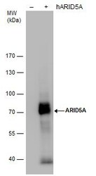
- ARID5A Polyclonal Antibody detects ARID5A protein by western blot analysis. Non-transfected (-) and ARID5A-transfected (+, including 3xFlag-tag) 293T whole cell extracts (30 µg) were separated by 7.5% SDS-PAGE, and the membrane was blotted with ARID5A Polyclonal Antibody ARID5A Monoclonal Antibody (P18112) (Product # MA5-18292) diluted by 1:5,000.
- Submitted by
- Invitrogen Antibodies (provider)
- Main image
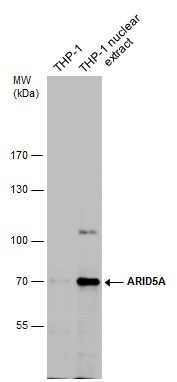
- Experimental details
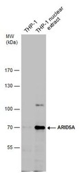
- Western Blot analysis of ARID5A was performed by separating 30 µg of THP-1 whole cell and nuclear extracts by 7.5% SDS-PAGE. Proteins were transferred to a membrane and probed with a ARID5A Monoclonal Antibody (P18112) (Product # MA5-18292) at a dilution of 1:500The signal was developed with Trident ECL plus-Enhanced.
- Submitted by
- Invitrogen Antibodies (provider)
- Main image
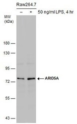
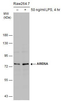
- Experimental details
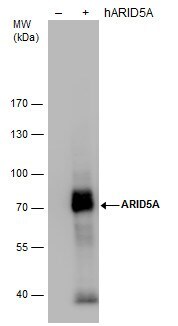
- Western Blot analysis of ARID5A was performed by separating 30 µg of untreated (–) and treated (+) Raw264.7 whole cell extracts by 5% SDS-PAGE. Proteins were transferred to a membrane and probed with a ARID5A Monoclonal Antibody (P18112) (Product # MA5-18292) at a dilution of 1:500. (LPS: Lipopolysaccharides).
Supportive validation
- Submitted by
- Invitrogen Antibodies (provider)
- Main image
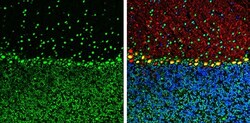
- Experimental details
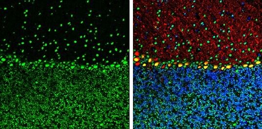
- Immunohistochemistry (Frozen) analysis of ARID5A was performed in frozen-sectioned mouse mouse cerebellum tissue using ARID5A Monoclonal Antibody (P18112) (Product # MA5-18292) at a dilution of 1:250 (Green). Red: Calbindin, stained by Calbindin antibody diluted at 1:500. Blue: Fluoroshield with DAPI. Antigen Retrieval: Citrate buffer, pH 6.0, 10 min.
Supportive validation
- Submitted by
- Invitrogen Antibodies (provider)
- Main image

- Experimental details
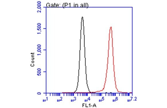
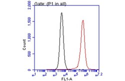
- Flow Cytometry analysis of ARID5A was performed in THP-1 cells using ARID5A Monoclonal Antibody (P18112) (Product # MA5-18292) (red) at a dilution of 1:20. Black: Unlabelled sample was used as a control. Acquisition of 20,000 events were collected using a Dylight 488-conjugated secondary antibody for FACS analysis.

- Submitted by
- Invitrogen Antibodies (provider)
- Main image
- Experimental details
- Flow Cytometry analysis of ARID5A was performed in THP-1 cells using ARID5A Monoclonal Antibody (P18112) (Product # MA5-18292) (red) at a dilution of 1:20. Black: Unlabelled sample was used as a control. Acquisition of 20,000 events were collected using a Dylight 488-conjugated secondary antibody for FACS analysis.