Explore
 Validate
 Learn
Learn Western blot
 ELISA
ELISAAntibody data
- Antibody Data
- Antigen structure
- References [3]
- Comments [0]
- Validations
- Western blot [1]
- Immunohistochemistry [1]
- Blocking/Neutralizing [1]
Submit
Validation data
Reference
Comment
Report error
- Product number
- AF682 - Provider product page

- Provider
- R&D Systems
- Product name
- Human Prolactin Antibody
- Antibody type
- Polyclonal
- Description
- Antigen Affinity-purified. Detects human Prolactin in ELISAs and Western blots. In sandwich ELISAs, less than 0.05% cross-reactivity with recombinant mouse Prolactin and recombinant human Prolactin R.
- Reactivity
- Human
- Host
- Goat
- Conjugate
- Unconjugated
- Antigen sequence
Q5THQ0- Isotype
- IgG
- Vial size
- 100 ug
- Concentration
- LYOPH
- Storage
- Use a manual defrost freezer and avoid repeated freeze-thaw cycles. 12 months from date of receipt, -20 to -70 °C as supplied. 1 month, 2 to 8 °C under sterile conditions after reconstitution. 6 months, -20 to -70 °C under sterile conditions after reconstitution.
Submitted references Prolactin enhances interferon-gamma-induced production of CXC ligand 9 (CXCL9), CXCL10, and CXCL11 in human keratinocytes.
Shift of monocyte function toward cellular immunity during sleep.
Prolactin and heregulin override DNA damage-induced growth arrest and promote phosphatidylinositol-3 kinase-dependent proliferation in breast cancer cells.
Kanda N, Watanabe S
Endocrinology 2007 May;148(5):2317-25
Endocrinology 2007 May;148(5):2317-25
Shift of monocyte function toward cellular immunity during sleep.
Lange T, Dimitrov S, Fehm HL, Westermann J, Born J
Archives of internal medicine 2006 Sep 18;166(16):1695-700
Archives of internal medicine 2006 Sep 18;166(16):1695-700
Prolactin and heregulin override DNA damage-induced growth arrest and promote phosphatidylinositol-3 kinase-dependent proliferation in breast cancer cells.
Chakravarti P, Henry MK, Quelle FW
International journal of oncology 2005 Feb;26(2):509-14
International journal of oncology 2005 Feb;26(2):509-14
No comments: Submit comment
Supportive validation
- Submitted by
- R&D Systems (provider)
- Main image
- Experimental details
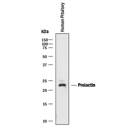
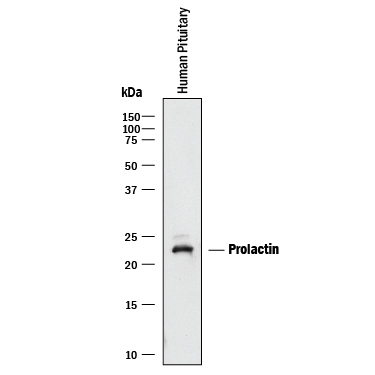
- Detection of Human Prolactin by Western Blot. Western blot shows lysates of human pituitary tissue. PVDF membrane was probed with 0.25 µg/mL of Goat Anti-Human Prolactin Antigen Affinity-purified Polyclonal Antibody (Catalog # AF682) followed by HRP-conjugated Anti-Goat IgG Secondary Antibody (Catalog # HAF017). A specific band was detected for Prolactin at approximately 23 kDa (as indicated). This experiment was conducted under reducing conditions and using Immunoblot Buffer Group 1.
Supportive validation
- Submitted by
- R&D Systems (provider)
- Main image

- Experimental details
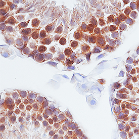
- Prolactin in Human Testis. Prolactin was detected in immersion fixed paraffin-embedded sections of human testis using Goat Anti-Human Prolactin Antigen Affinity-purified Polyclonal Antibody (Catalog # AF682) at 1 µg/mL overnight at 4 °C. Tissue was stained using the Anti-Goat HRP-DAB Cell & Tissue Staining Kit (brown; Catalog # CTS008) and counterstained with hematoxylin (blue). Specific staining was localized to cytoplasm of sperm cells. View our protocol for Chromogenic IHC Staining of Paraffin-embedded Tissue Sections.
Supportive validation
- Submitted by
- R&D Systems (provider)
- Main image
- Experimental details
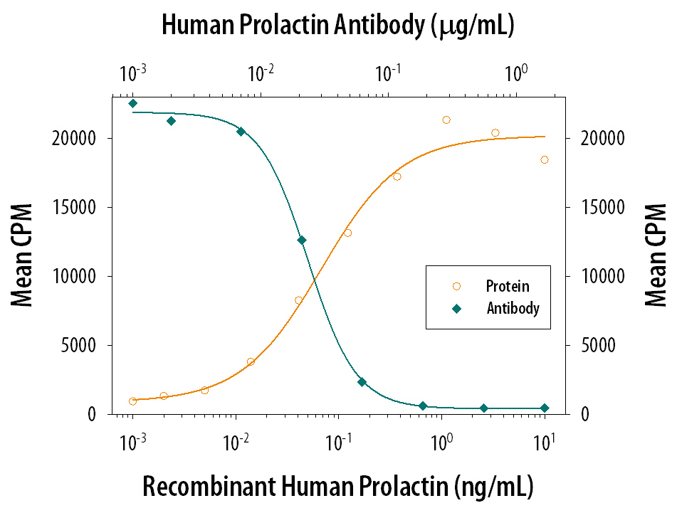
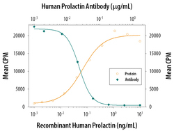
- Cell Proliferation Induced by Prolactin and Neutralization by Human Prolactin Antibody. Recombinant Human Prolactin (Catalog # 682-PL) stimulates proliferation in the Nb2-11 rat lymphoma cell line in a dose-dependent manner (orange line). Proliferation elicited by Recombinant Human Prolactin (0.5 ng/mL) is neutralized (green line) by increasing concentrations of Goat Anti-Human Prolactin Antigen Affinity-purified Polyclonal Antibody (Catalog # AF682). The ND50 is typically 0.02-0.05 µg/mL.