Explore
 Validate
 Learn
Learn Western blot
Antibody data
- Antibody Data
- Antigen structure
- References [1]
- Comments [0]
- Validations
- Western blot [3]
- Immunocytochemistry [3]
- Flow cytometry [1]
Submit
Validation data
Reference
Comment
Report error
- Product number
- PA1-880 - Provider product page

- Provider
- Invitrogen Antibodies
- Product name
- DNMT1 Polyclonal Antibody
- Antibody type
- Polyclonal
- Antigen
- Synthetic peptide
- Description
- PA1-880 detects DNA methyltransferase 1 (Dmnt1) from HeLa cell nuclear extracts. PA1-880 has been successfully used in Western blot procedures. By Western blot, this antibody detects an ~180 kDa protein representing Dnmt1 from HeLa cell nuclear extract. A doublet between 40 and 45 kDa is also seen on Western blots using this antibody. The PA1-880 immunogen is a synthetic peptide corresponding to residues A(171) K G P A K R K P Q E E S E(185) of human Dnmt1. PA1-880 immunizing peptide (Cat. # PEP-136) is available for use in neutralization and control experiments.
- Reactivity
- Human, Rat
- Host
- Rabbit
- Isotype
- IgG
- Vial size
- 100 µg
- Concentration
- 1 mg/mL
- Storage
- -20° C, Avoid Freeze/Thaw Cycles
Submitted references Expression of an alternative Dnmt1 isoform during muscle differentiation.
Aguirre-Arteta AM, Grunewald I, Cardoso MC, Leonhardt H
Cell growth & differentiation : the molecular biology journal of the American Association for Cancer Research 2000 Oct;11(10):551-9
Cell growth & differentiation : the molecular biology journal of the American Association for Cancer Research 2000 Oct;11(10):551-9
No comments: Submit comment
Supportive validation
- Submitted by
- Invitrogen Antibodies (provider)
- Main image
- Experimental details
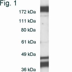
- Western blot of Dnmt1 on HeLa cell nuclear extract using Product # PA1-880.
- Submitted by
- Invitrogen Antibodies (provider)
- Main image

- Experimental details
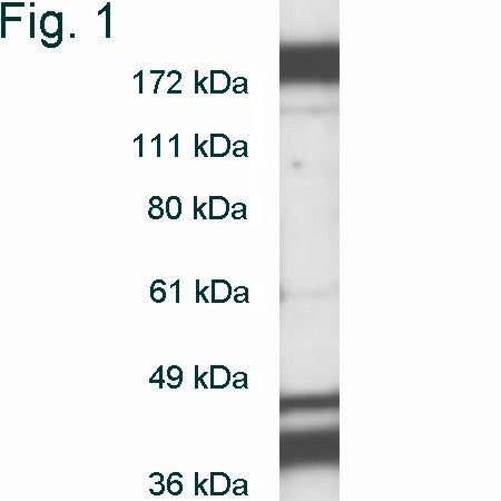
- Western blot of Dnmt1 on HeLa cell nuclear extract using Product # PA1-880.
- Submitted by
- Invitrogen Antibodies (provider)
- Main image
- Experimental details
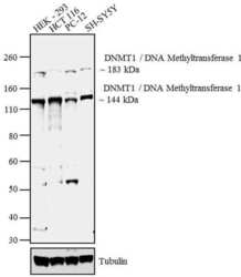
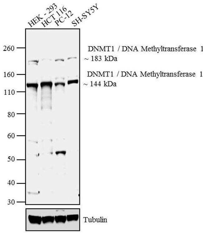
- Western blot analysis was performed nuclear enriched lysates of HEK -293 (Lane 1), HCT 116 (Lane2), PC-12 (Lane 3) and SH-SY5Y (Lane 4). The blots were probed with Anti- DNMT1/DNA Methyltransferase 1 Rabbit Polyclonal Antibody (Product # PA1-880, 2 µg/mL) and detected by chemiluminescence using Goat anti-Rabbit IgG (H+L) Superclonal™ Secondary Antibody, HRP conjugate (Product # A27036, 0.4 µg/mL, 1:2500 dilution). Bands of ~183 and ~144 kDa corresponding to DNMT1/DNA Methyltransferase 1 were observed across the cell lines tested. Known quantity of protein samples were electrophoresed using Novex® NuPAGE® 4-12 % Bis-Tris gel (Product # NP0321BOX), XCell SureLock™ Electrophoresis System (Product # EI0002) and Novex® Sharp Pre-Stained Protein Standard (Product # LC5800). Resolved proteins were then transferred onto a nitrocellulose membrane by overnight transfer method. The membrane was probed with the relevant primary and secondary Antibody following blocking with 5 % skimmed milk. Chemiluminescent detection was performed using Pierce™ ECL Western Blotting Substrate (Product # 32106).
Supportive validation
- Submitted by
- Invitrogen Antibodies (provider)
- Main image
- Experimental details
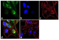
- Immunofluorescent analysis of DNMT1/DNA Methyltransferase 1 was performed using 70% confluent log phase PANC-1 cells treated with 100 ng of Interleukin-6 for 30 minutes. The cells were fixed with 4% paraformaldehyde for 10 minutes, permeabilized with 0.1% Triton™ X-100 for 10 minutes, and blocked with 1% BSA for 1 hour at room temperature. The cells were labeled with DNMT1/DNA Methyltransferase 1 Rabbit Polyclonal Antibody (Product # PA1-880) at 2 µg/mL in 0.1% BSA and incubated for 3 hours at room temperature and then labeled with Goat anti-Rabbit IgG (H+L) Superclonal™ Secondary Antibody, Alexa Fluor® 488 conjugate (Product # A27034) a dilution of 1:2000 for 45 minutes at room temperature (Panel a: green). Nuclei (Panel b: blue) were stained with SlowFade® Gold Antifade Mountant with DAPI (Product # S36938). F-actin (Panel c: red) was stained with Alexa Fluor® 555 Rhodamine Phalloidin (Product # R415, 1:300). Panel d represents the merged image showing predominantly nuclear localization. Panel e is untreated cell with predominantly cytoplasmic signal. Panel f represents control cells with no primary antibody to assess background. The images were captured at 60X magnification.

- Submitted by
- Invitrogen Antibodies (provider)
- Main image
- Experimental details
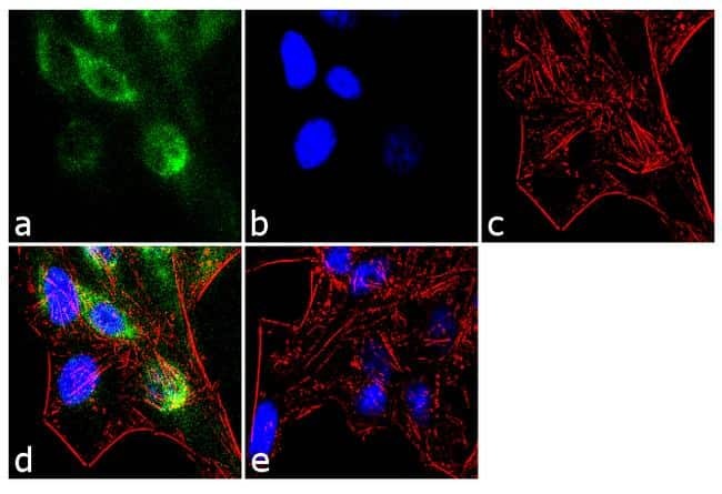
- Immunofluorescent analysis of DNMT1/DNA Methyltransferase 1 was performed using 70% confluent log phase HeLa cells. The cells were fixed with 4% paraformaldehyde for 10 minutes, permeabilized with 0.1% Triton™ X-100 for 10 minutes, and blocked with 1% BSA for 1 hour at room temperature. The cells were labeled with DNMT1/DNA Methyltransferase 1 Rabbit Polyclonal Antibody (Product # PA1-880) at 2 µg/mL in 0.1% BSA and incubated for 3 hours at room temperature and then labeled with Goat anti-Rabbit IgG (H+L) Superclonal™ Secondary Antibody, Alexa Fluor® 488 conjugate (Product # A27034) a dilution of 1:2000 for 45 minutes at room temperature (Panel a: green). Nuclei (Panel b: blue) were stained with SlowFade® Gold Antifade Mountant with DAPI (Product # S36938). F-actin (Panel c: red) was stained with Alexa Fluor® 555 Rhodamine Phalloidin (Product # R415, 1:300). Panel d represents the merged image showing predominantly cytoplasmic localization. Panel e shows the no primary antibody control. The images were captured at 60X magnification.
- Submitted by
- Invitrogen Antibodies (provider)
- Main image
- Experimental details
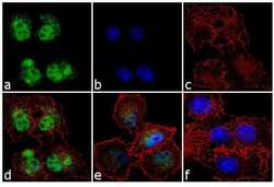
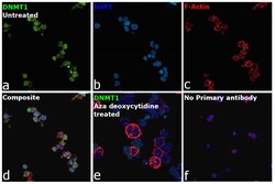
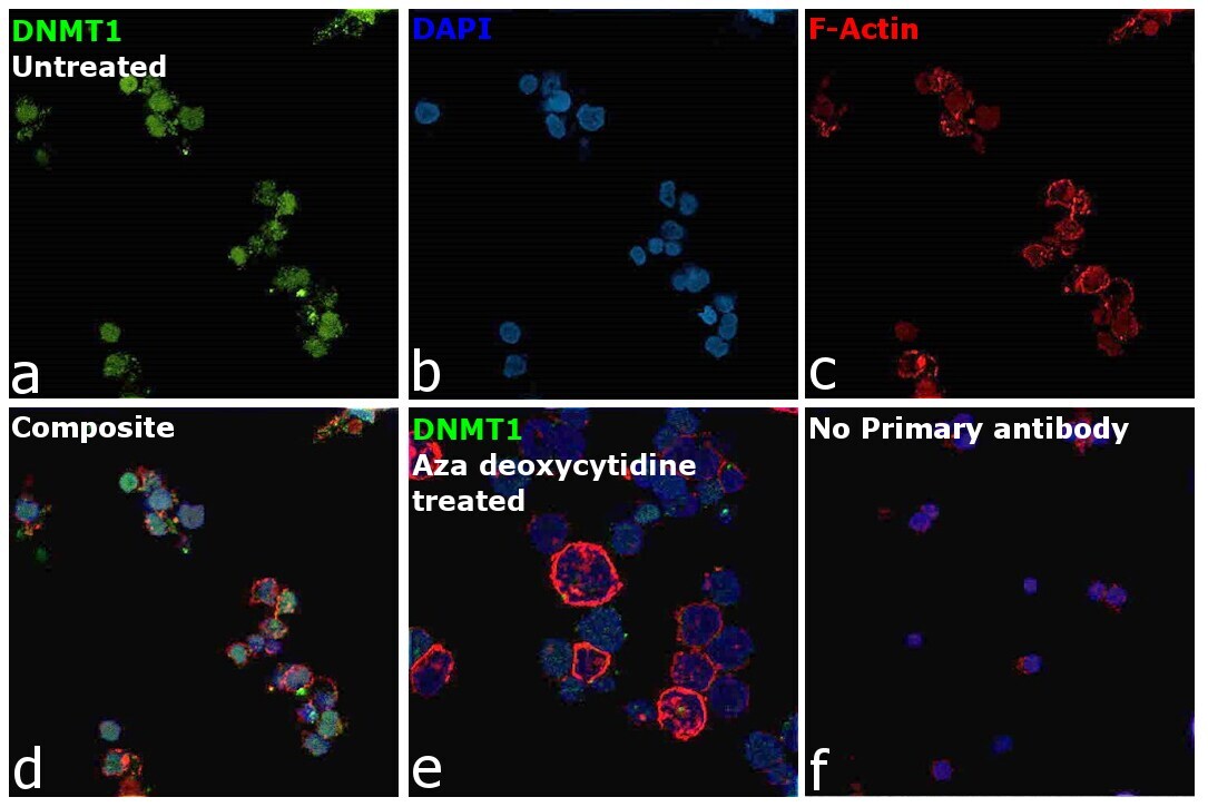
- Immunofluorescence analysis of DNMT1 was performed using 70% confluent log phase K-562 cells. The cells were fixed with 4% paraformaldehyde for 10 minutes, permeabilized with 0.1% Triton™ X-100 for 15 minutes, and blocked with 2% BSA for 1 hour at room temperature. The cells were labeled with DNMT1 Polyclonal Antibody (Product # PA1-880) at 2 µg/mL in 0.1% BSA, incubated at 4 degree celsius overnight and then labeled with Donkey anti-Rabbit IgG (H+L) Highly Cross-Adsorbed Secondary Antibody, Alexa Fluor Plus 488 (Product # A32790), (1:2000), for 45 minutes at room temperature (Panel a: Green). Nuclei (Panel b:Blue) were stained with ProLong™ Diamond Antifade Mountant with DAPI (Product # P36962). F-actin (Panel c: Red) was stained with Rhodamine Phalloidin (Product # R415, 1:300). Panel d represents the merged image showing predominant nuclear localization. Panel e represents merged image of K-562 cells treated with 5 µM Aza deoxycytidine for 3 days (medium and drug were replaced every 24 hours). The signal is significantly lower in panel e due to down-regulation of DNMT1 expression upon Aza deoxycytidine treatment. Panel f represents untreated control cells with no primary antibody to assess background. The images were captured at 60X magnification.
Supportive validation
- Submitted by
- Invitrogen Antibodies (provider)
- Main image

- Experimental details
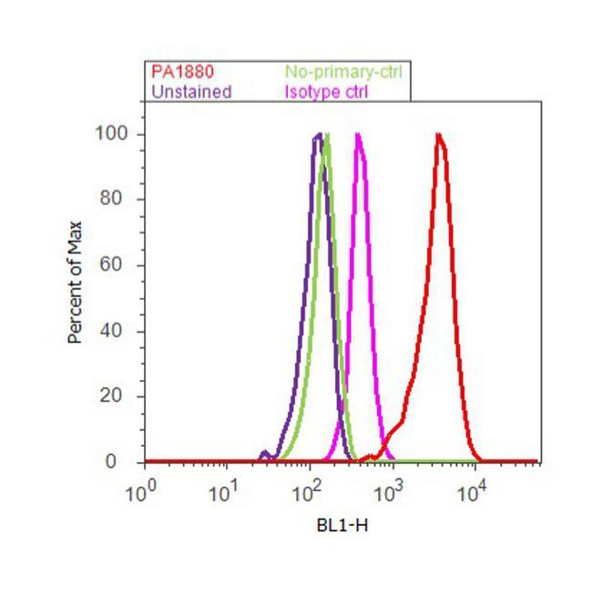
- Flow cytometry analysis of DNMT1/DNA Methyltransferase 1 was done on HCT 116 cells. Cells were fixed with 70% ethanol for 10 minutes, permeabilized with 0.25% Triton™ X-100 for 20 minutes, and blocked with 5% BSA for 30 minutes at room temperature. Cells were labeled with DNMT1 Rabbit Polyclonal Antibody (PA1-880, red histogram) or with rabbit isotype control (pink histogram) at 3-5 ug/million cells in 2.5% BSA. After incubation at room temperature for 2 hours, the cells were labeled with Alexa Fluor® 488 Goat Anti-Rabbit Secondary Antibody (A11008) at a dilution of 1:400 for 30 minutes at room temperature. The representative 10, 000 cells were acquired and analyzed for each sample using an Attune® Acoustic Focusing Cytometer. The purple histogram represents unstained control cells and the green histogram represents no-primary-antibody control.