Explore
 Validate
 Learn
Learn Western blot
 Immunocytochemistry
Antibody data
- Antibody Data
- Antigen structure
- References [0]
- Comments [0]
- Validations
- Immunocytochemistry [7]
- Immunoprecipitation [1]
- Immunohistochemistry [3]
- Chromatin Immunoprecipitation [4]
- Other assay [1]
Submit
Validation data
Reference
Comment
Report error
- Product number
- PA5-30581 - Provider product page

- Provider
- Invitrogen Antibodies
- Product name
- DNMT1 Polyclonal Antibody
- Antibody type
- Polyclonal
- Antigen
- Synthetic peptide
- Description
- Recommended positive controls: 293T, A431, HeLa, HepG2, Jurkat, Raji, NCI-H929. Store product as a concentrated solution. Centrifuge briefly prior to opening the vial.
- Reactivity
- Human, Mouse
- Host
- Rabbit
- Isotype
- IgG
- Vial size
- 100 μL
- Concentration
- 0.18 mg/mL
- Storage
- Store at 4°C short term. For long term storage, store at -20°C, avoiding freeze/thaw cycles.
No comments: Submit comment
Supportive validation
- Submitted by
- Invitrogen Antibodies (provider)
- Main image
- Experimental details
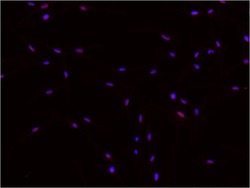
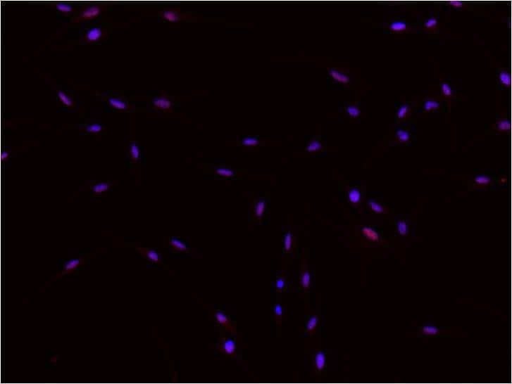
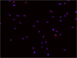
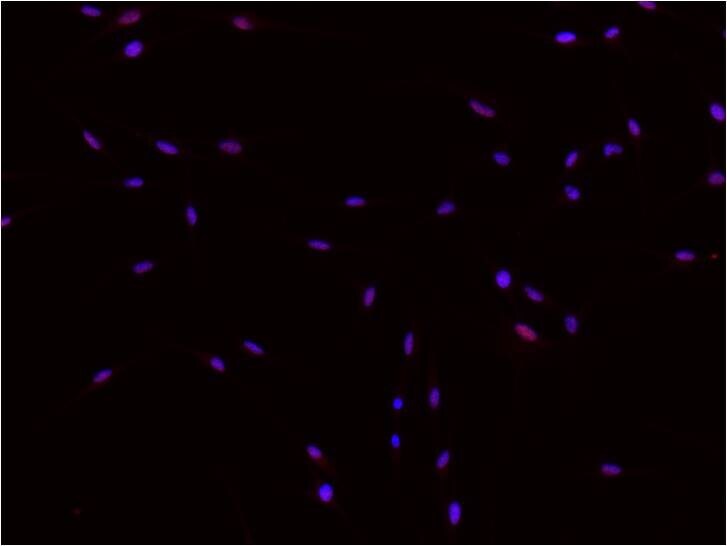
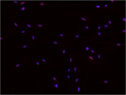
- Immunofluorescent analysis of DNMT1 (red) in human melanocytes. Cells were fixed with 4% paraformaldehyde for 15 minutes, permeabilized with 0.2% Triton X-100 in PBS for 10 minutes at room temperature, and blocked with 5% normal goat serum for 1 hour at room temperature. Cells were probed with a DNMT1 polyclonal antibody (Product # PA5-30581) at a dilution of 1:500 for 1 hour at room temperature, washed with PBS, and incubated with an anti-rabbit Alexa-Fluor 555-conjugated secondary antibody at a dilution of 1:500 for 1 hour at room temperature. Nuclei (blue) were stained with DAPI. Images were taken on a fluorescent microscope at 20X magnification. Data courtesy of the Innovators Program.
- Submitted by
- Invitrogen Antibodies (provider)
- Main image
- Experimental details
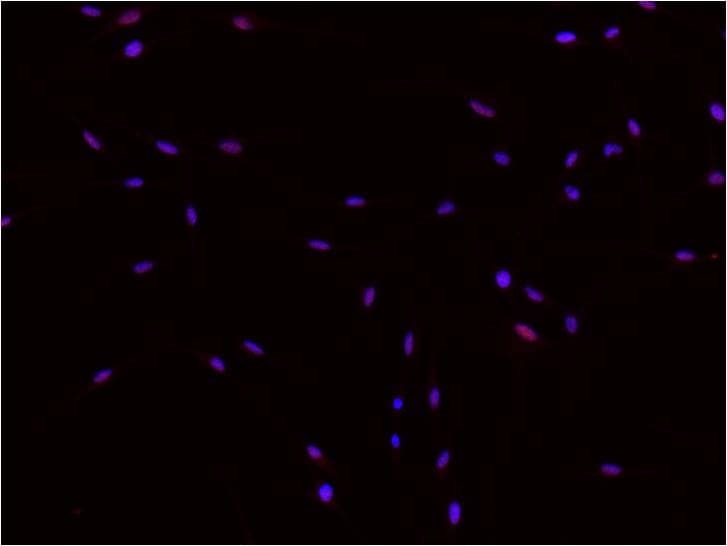
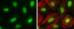
- Immunocytochemistry-Immunofluorescence analysis of DNMT1 was performed in HeLa cells fixed in 4% paraformaldehyde at RT for 15 min. Green: DNMT1 Polyclonal Antibody (Product # PA5-30581) diluted at 1:2000. Red: phalloidin, a cytoskeleton marker.
- Submitted by
- Invitrogen Antibodies (provider)
- Main image
- Experimental details
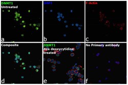
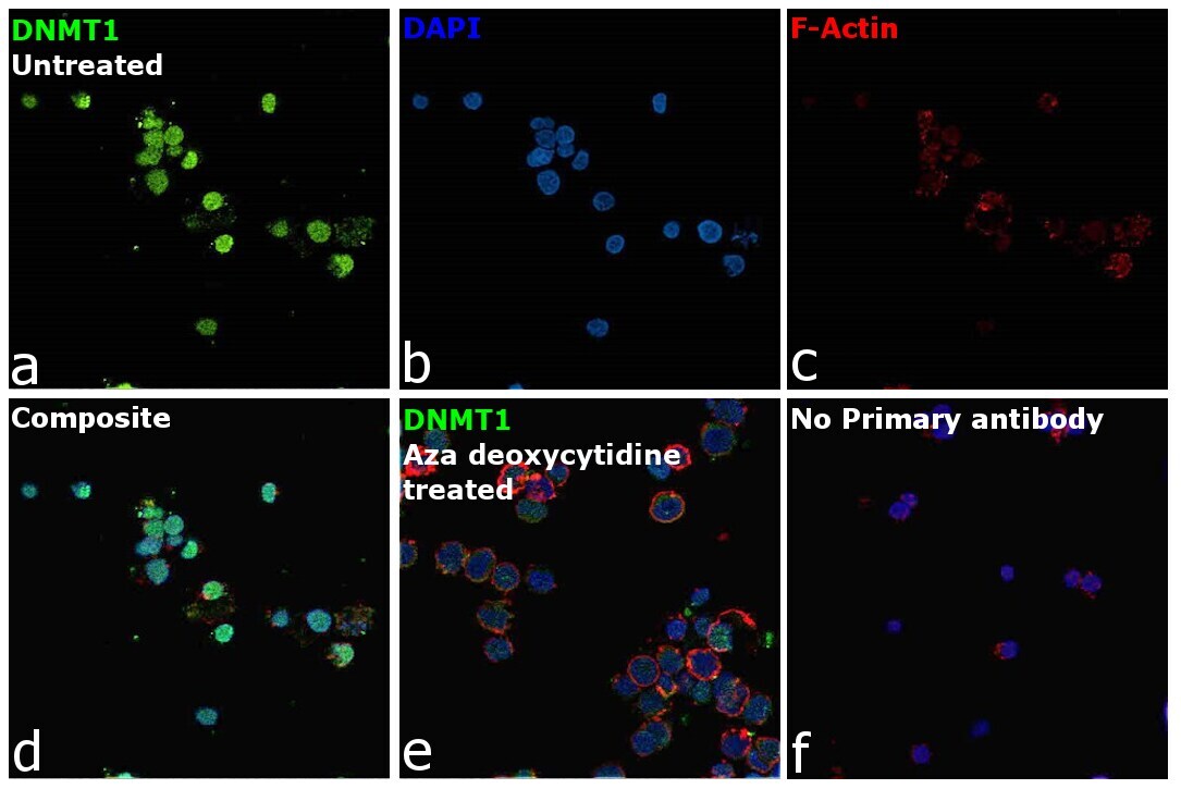
- Immunofluorescence analysis of DNMT1 was performed using 70% confluent log phase K-562 cells. The cells were fixed with 4% paraformaldehyde for 10 minutes, permeabilized with 0.1% Triton™ X-100 for 15 minutes, and blocked with 2% BSA for 1 hour at room temperature. The cells were labeled with DNMT1 Polyclonal Antibody (Product # PA5-30581) at 1:500 in 0.1% BSA, incubated at 4 degree celsius overnight and then labeled with Donkey anti-Rabbit IgG (H+L) Highly Cross-Adsorbed Secondary Antibody, Alexa Fluor Plus 488 (Product # A32790), (1:2000), for 45 minutes at room temperature (Panel a: Green). Nuclei (Panel b:Blue) were stained with ProLong™ Diamond Antifade Mountant with DAPI (Product # P36962). F-actin (Panel c: Red) was stained with Rhodamine Phalloidin (Product # R415, 1:300). Panel d represents the merged image showing predominant nuclear localization. Panel e represents merged image of K-562 cells treated with 5 µM Aza deoxycytidine for 3 days (medium and drug were replaced every 24 hours). The signal is significantly lower in panel e due to down-regulation of DNMT1 expression upon Aza deoxycytidine treatment. Panel f represents untreated control cells with no primary antibody to assess background. The images were captured at 60X magnification.
- Submitted by
- Invitrogen Antibodies (provider)
- Main image

- Experimental details
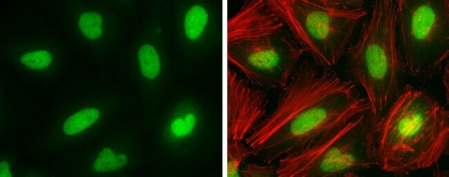
- Immunocytochemistry-Immunofluorescence analysis of DNMT1 was performed in HeLa cells fixed in 4% paraformaldehyde at RT for 15 min. Green: DNMT1 Polyclonal Antibody (Product # PA5-30581) diluted at 1:2000. Red: phalloidin, a cytoskeleton marker.

- Submitted by
- Invitrogen Antibodies (provider)
- Main image

- Experimental details
- Immunofluorescence analysis of DNMT1 was performed using 70% confluent log phase K-562 cells. The cells were fixed with 4% paraformaldehyde for 10 minutes, permeabilized with 0.1% Triton™ X-100 for 15 minutes, and blocked with 2% BSA for 1 hour at room temperature. The cells were labeled with DNMT1 Polyclonal Antibody (Product # PA5-30581) at 1:500 in 0.1% BSA, incubated at 4 degree celsius overnight and then labeled with Donkey anti-Rabbit IgG (H+L) Highly Cross-Adsorbed Secondary Antibody, Alexa Fluor Plus 488 (Product # A32790), (1:2000), for 45 minutes at room temperature (Panel a: Green). Nuclei (Panel b:Blue) were stained with ProLong™ Diamond Antifade Mountant with DAPI (Product # P36962). F-actin (Panel c: Red) was stained with Rhodamine Phalloidin (Product # R415, 1:300). Panel d represents the merged image showing predominant nuclear localization. Panel e represents merged image of K-562 cells treated with 5 µM Aza deoxycytidine for 3 days (medium and drug were replaced every 24 hours). The signal is significantly lower in panel e due to down-regulation of DNMT1 expression upon Aza deoxycytidine treatment. Panel f represents untreated control cells with no primary antibody to assess background. The images were captured at 60X magnification.

- Submitted by
- Invitrogen Antibodies (provider)
- Main image
- Experimental details
- Immunofluorescent analysis of DNMT1 (red) in human melanocytes. Cells were fixed with 4% paraformaldehyde for 15 minutes, permeabilized with 0.2% Triton X-100 in PBS for 10 minutes at room temperature, and blocked with 5% normal goat serum for 1 hour at room temperature. Cells were probed with a DNMT1 polyclonal antibody (Product # PA5-30581) at a dilution of 1:500 for 1 hour at room temperature, washed with PBS, and incubated with an anti-rabbit Alexa-Fluor 555-conjugated secondary antibody at a dilution of 1:500 for 1 hour at room temperature. Nuclei (blue) were stained with DAPI. Images were taken on a fluorescent microscope at 20X magnification. Data courtesy of the Innovators Program.
Supportive validation
- Submitted by
- Invitrogen Antibodies (provider)
- Main image

- Experimental details
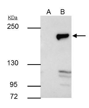
- DNMT1 Polyclonal Antibody immunoprecipitates DNMT1 protein in IP experiments. IP samples: HeLa nuclear extract. A. Control with 4 µg of preimmune Rabbit IgG. B. Immunoprecipitation of DNMT1 protein by 4 µg DNMT1 Polyclonal Antibody (Product # PA5-30581). 5 % SDS-PAGE. The immunoprecipitated DNMT1 protein was detected by DNMT1 Polyclonal Antibody (Product # PA5-30581) diluted at 1:1,000.

Supportive validation
- Submitted by
- Invitrogen Antibodies (provider)
- Main image
- Experimental details
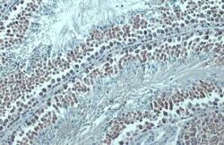
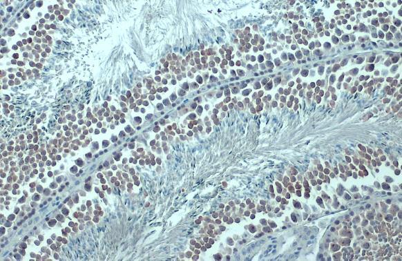
- DNMT1 Polyclonal Antibody detects DNMT1 protein at cytoplasm by immunohistochemical analysis. Sample: Paraffin-embedded mouse testis. DNMT1 stained by DNMT1 Polyclonal Antibody (Product # PA5-30581) diluted at 1:500. Antigen Retrieval: Citrate buffer, pH 6.0, 15 min.
- Submitted by
- Invitrogen Antibodies (provider)
- Main image
- Experimental details
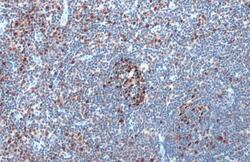
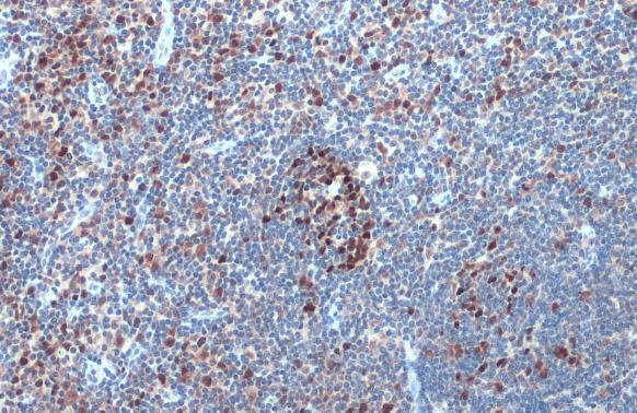
- DNMT1 Polyclonal Antibody detects DNMT1 protein at nucleus by immunohistochemical analysis. Sample: Paraffin-embedded human tonsil. DNMT1 stained by DNMT1 Polyclonal Antibody (Product # PA5-30581) diluted at 1:500. Antigen Retrieval: Citrate buffer, pH 6.0, 15 min.
- Submitted by
- Invitrogen Antibodies (provider)
- Main image
- Experimental details
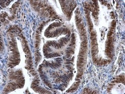
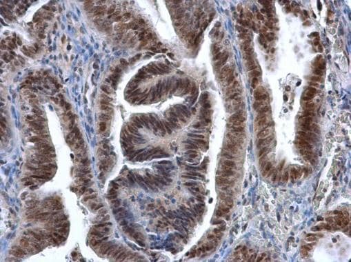
- Immunohistochemistry (Paraffin) analysis of DNMT1 was performed in paraffin-embedded human endometrial cancer tissue using DNMT1 Polyclonal Antibody (Product # PA5-30581) at a dilution of 1:500.
Supportive validation
- Submitted by
- Invitrogen Antibodies (provider)
- Main image
- Experimental details
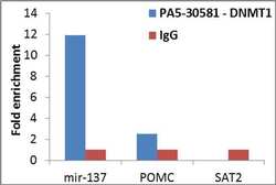
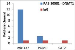
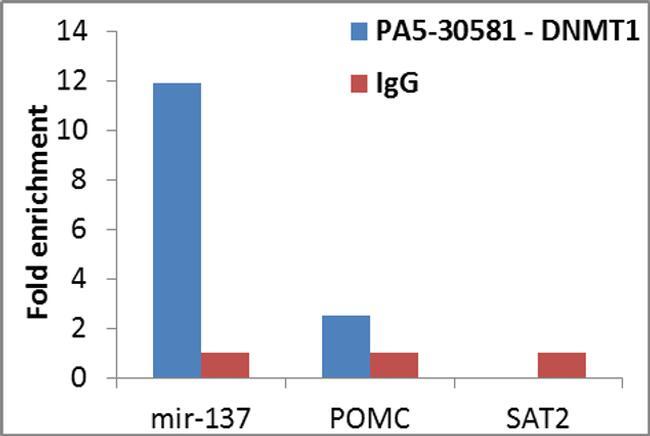
- Enrichment of endogenous DNMT1 protein at specific gene loci using Anti-DNMT1 Antibody: Chromatin Immunoprecipitation (ChIP) was performed using Anti-DNMT1 Rabbit Polyclonal Antibody (Product # PA5-30581, 4 ug) on sheared chromatin from 2 million HeLa cells using the MAGnify ChIP System (Product # 49-2024). Normal Rabbit IgG was used as a negative IP control. The purified DNA was analyzed by qPCR with PCR primer pairs over mir-137 and POMC (positive) and SAT2 satellite repeats (negative). Data is presented as fold enrichment of the antibody signal versus the negative control IgG using the comparative CT method.
- Submitted by
- Invitrogen Antibodies (provider)
- Main image
- Experimental details
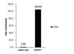
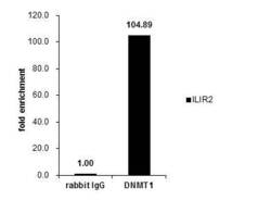
- Cross-linked ChIP was performed with HCT116 chromatin extract and 5 µg of either control rabbit IgG or DNMT1 Polyclonal Antibody (Product # PA5-30581). The precipitated DNA was detected by PCR with primer set targeting to ILIR2.

- Submitted by
- Invitrogen Antibodies (provider)
- Main image
- Experimental details
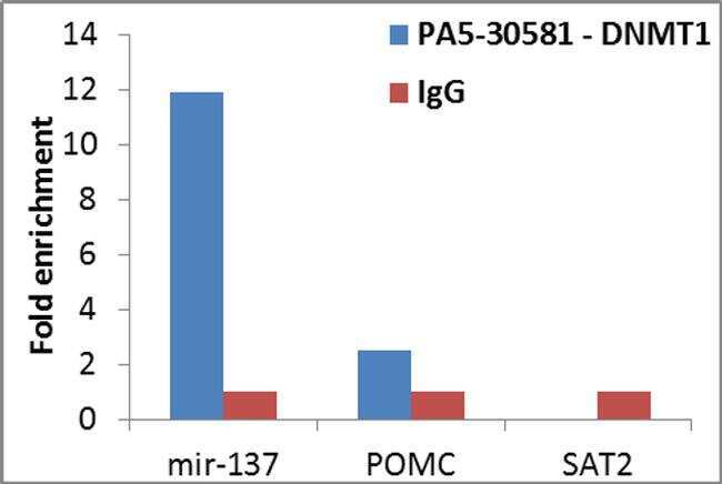
- Enrichment of endogenous DNMT1 protein at specific gene loci using Anti-DNMT1 Antibody: Chromatin Immunoprecipitation (ChIP) was performed using Anti-DNMT1 Rabbit Polyclonal Antibody (Product # PA5-30581, 4 ug) on sheared chromatin from 2 million HeLa cells using the MAGnify ChIP System (Product # 49-2024). Normal Rabbit IgG was used as a negative IP control. The purified DNA was analyzed by qPCR with PCR primer pairs over mir-137 and POMC (positive) and SAT2 satellite repeats (negative). Data is presented as fold enrichment of the antibody signal versus the negative control IgG using the comparative CT method.
- Submitted by
- Invitrogen Antibodies (provider)
- Main image
- Experimental details
- Cross-linked ChIP was performed with HCT116 chromatin extract and 5 µg of either control rabbit IgG or DNMT1 Polyclonal Antibody (Product # PA5-30581). The precipitated DNA was detected by PCR with primer set targeting to ILIR2.

Supportive validation
- Submitted by
- Invitrogen Antibodies (provider)
- Main image

- Experimental details
- DNMT1 Polyclonal Antibody immunoprecipitates DNMT1 protein in IP experiments. IP samples: HeLa nuclear extract. A. Control with 4 µg of preimmune Rabbit IgG. B. Immunoprecipitation of DNMT1 protein by 4 µg DNMT1 Polyclonal Antibody (Product # PA5-30581). 5 % SDS-PAGE. The immunoprecipitated DNMT1 protein was detected by DNMT1 Polyclonal Antibody (Product # PA5-30581) diluted at 1:1,000.