 Explore
 Validate
 Learn
Learn Western blot
Antibody data
- Antibody Data
- Antigen structure
- References [0]
- Comments [0]
- Validations
- Western blot [2]
- Immunocytochemistry [2]
Submit
Validation data
Reference
Comment
Report error
- Product number
- AF6110 - Provider product page

- Provider
- R&D Systems
- Product name
- Human DNMT1 Antibody
- Antibody type
- Polyclonal
- Description
- Antigen Affinity-purified. Detects human DNMT1 in Western blots.
- Reactivity
- Human
- Host
- Sheep
- Conjugate
- Unconjugated
- Antigen sequence
P26358- Isotype
- IgG
- Vial size
- 100 ug
- Concentration
- LYOPH
- Storage
- Use a manual defrost freezer and avoid repeated freeze-thaw cycles. 12 months from date of receipt, -20 to -70 °C as supplied. 1 month, 2 to 8 °C under sterile conditions after reconstitution. 6 months, -20 to -70 °C under sterile conditions after reconstitution.
No comments: Submit comment
Supportive validation
- Submitted by
- R&D Systems (provider)
- Main image
- Experimental details
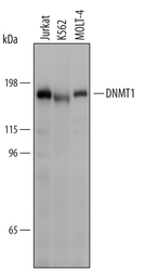
- Detection of Human DNMT1 by Western Blot. Western blot shows lysates of Jurkat human acute T cell leukemia cell line, K562 human chronic myelogenous leukemia cell line, and MOLT-4 human acute lymphoblastic leukemia cell line. PVDF Membrane was probed with 1 µg/mL of Sheep Anti-Human DNMT1 Antigen Affinity-purified Polyclonal Antibody (Catalog # AF6110) followed by HRP-conjugated Anti-Sheep IgG Secondary Antibody (Catalog # HAF016). A specific band was detected for DNMT1 at approximately 183 kDa (as indicated). This experiment was conducted under reducing conditions and using Immunoblot Buffer Group 1.
- Submitted by
- R&D Systems (provider)
- Main image

- Experimental details
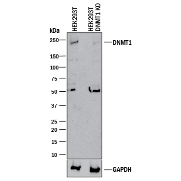
- Western Blot Shows Human DNMT1 Specificity by Using Knockout Cell Line. Western blot shows lysates of HEK293T human embryonic kidney parental cell line and DNMT1 knockout HEK293T cell line (KO). PVDF membrane was probed with 1 µg/mL of Sheep Anti-Human DNMT1 Antigen Affinity-purified Polyclonal Antibody (Catalog # AF6110) followed by HRP-conjugated Anti-Sheep IgG Secondary Antibody (Catalog # HAF016). A specific band was detected for DNMT1 at approximately 230 kDa (as indicated) in the parental HEK293T cell line, but is not detectable in knockout HEK293T cell line. GAPDH (Catalog # AF5718) is shown as a loading control. This experiment was conducted under reducing conditions and using Immunoblot Buffer Group 1.
Supportive validation
- Submitted by
- R&D Systems (provider)
- Main image
- Experimental details
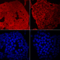
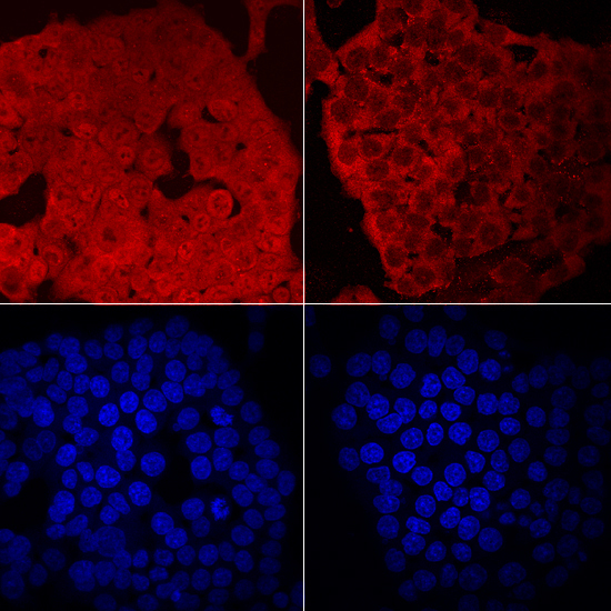
- DNMT1 in HCT-116 Human Cell Line. DNMT1 was detected in immersion fixed HCT-116 human colorectal carcinoma cell line untreated (left panels) and treated (right panels) with 1 μM 5-azacytidine for 24 hours using Sheep Anti-Human DNMT1 Antigen Affinity-purified Polyclonal Antibody (Catalog # AF6110) at 10 µg/mL for 3 hours at room temperature. Cells were stained using the NorthernLights™ 557-conjugated Anti-Sheep IgG Secondary Antibody (red, upper panels; Catalog # NL010) and counterstained with DAPI (blue, lower panels). Specific staining was localized to nuclei and cytoplasm. Nuclear staining was reduced following treatment with 5-azacytidine. View our protocol for Fluorescent ICC Staining of Cells on Coverslips.
- Submitted by
- R&D Systems (provider)
- Main image
- Experimental details
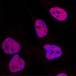
- DNMT1 in HeLa Human Cell Line. DNMT1 was detected in immersion fixed HeLa human cervical epithelial carcinoma cell line using Sheep Anti-Human DNMT1 Antigen Affinity-purified Polyclonal Antibody (Catalog # AF6110) at 1.7 µg/mL for 3 hours at room temperature. Cells were stained using the NorthernLights™ 557-conjugated Anti-Sheep IgG Secondary Antibody (red; Catalog # NL010) and counterstained with DAPI (blue). Specific staining was localized to nuclei. View our protocol for Fluorescent ICC Staining of Cells on Coverslips.
