Explore
 Validate
 Learn
Learn Western blot
Antibody data
- Antibody Data
- Antigen structure
- References [0]
- Comments [0]
- Validations
- Western blot [3]
- Immunohistochemistry [1]
- Flow cytometry [1]
Submit
Validation data
Reference
Comment
Report error
- Product number
- MAB9369-100 - Provider product page

- Provider
- R&D Systems
- Product name
- Human FANCD2 Antibody
- Antibody type
- Monoclonal
- Description
- Protein A or G purified from cell culture supernatant. Detects human FANCD2 in direct ELISAs and Western blots.
- Reactivity
- Human
- Host
- Rabbit
- Conjugate
- Unconjugated
- Antigen sequence
Q9BXW9- Isotype
- IgG
- Antibody clone number
- 1290D
- Vial size
- 100 ug
- Storage
- Use a manual defrost freezer and avoid repeated freeze-thaw cycles. 12 months from date of receipt, -20 to -70 °C as supplied. 1 month, 2 to 8 °C under sterile conditions after reconstitution. 6 months, -20 to -70 °C under sterile conditions after reconstitution.
No comments: Submit comment
Supportive validation
- Submitted by
- R&D Systems (provider)
- Main image

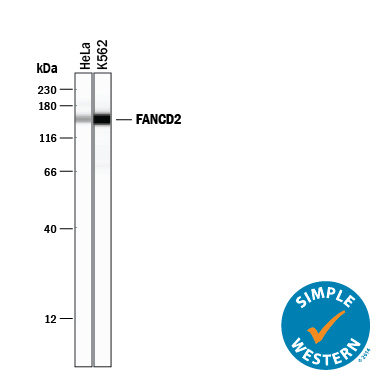
- Experimental details
- Detection of Human FANCD2 by Simple WesternTM. Simple Western lane view shows lysates of HeLa human cervical epithelial carcinoma cell line and K562 human chronic myelogenous leukemia cell line, loaded at 0.2 mg/mL. A specific band was detected for FANCD2 at approximately 152 kDa (as indicated) using 1 µg/mL of Rabbit Anti-Human FANCD2 Monoclonal Antibody (Catalog # MAB9369). This experiment was conducted under reducing conditions and using the 12-230 kDa separation system.
- Submitted by
- R&D Systems (provider)
- Main image
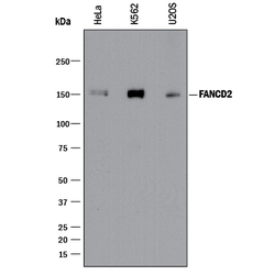
- Experimental details
- Detection of Human FANCD2 by Western Blot. Western blot shows lysates of HeLa human cervical epithelial carcinoma cell line, K562 human chronic myelogenous leukemia cell line, and U2OS human osteosarcoma cell line. PVDF membrane was probed with 0.1 µg/mL of Rabbit Anti-Human FANCD2 Monoclonal Antibody (Catalog # MAB9369) followed by HRP-conjugated Anti-Rabbit IgG Secondary Antibody (Catalog # HAF008). A specific band was detected for FANCD2 at approximately 150 kDa (as indicated). This experiment was conducted under reducing conditions and using Immunoblot Buffer Group 1.
- Submitted by
- R&D Systems (provider)
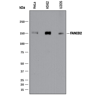
- Main image

- Experimental details
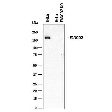
- Western Blot Shows Human FANCD2 Specificity Using Knockout Cell Line. Western blot shows lysates of HeLa human cervical epithelial carcinoma parental cell line and FANCD2 knockout (KO) HeLa cell line. PVDF membrane was probed with 0.1 µg/mL of Rabbit Anti-Human FANCD2 Monoclonal Antibody (Catalog # MAB9369) followed by HRP-conjugated Anti-Rabbit IgG Secondary Antibody (Catalog # HAF008). A specific band was detected for FANCD2 at approximately 150 kDa (as indicated) in the parental HeLa cell line, but is not detectable in the knockout HeLa cell line. This experiment was conducted under reducing conditions and using Immunoblot Buffer Group 1.
Supportive validation
- Submitted by
- R&D Systems (provider)
- Main image

- Experimental details
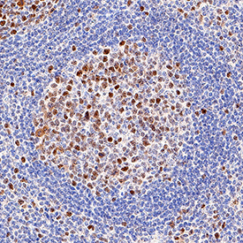
- FANCD2 in Human Tonsil. FANCD2 was detected in immersion fixed paraffin-embedded sections of human tonsil using Rabbit Anti-Human FANCD2 Monoclonal Antibody (Catalog # MAB9369) at 5 µg/mL for 1 hour at room temperature followed by incubation with the Anti-Rabbit IgG VisUCyte™ HRP Polymer Antibody (Catalog # VC003). Tissue was stained using DAB (brown) and counterstained with hematoxylin (blue). Specific staining was localized to nuclei. View our protocol for IHC Staining with VisUCyte HRP Polymer Detection Reagents.
Supportive validation
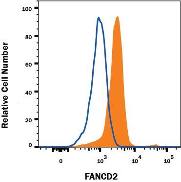
- Submitted by
- R&D Systems (provider)
- Main image
- Experimental details
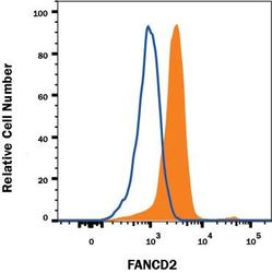
- Detection of FANCD2 in HeLa Human Cell Line by Flow Cytometry. HeLa human cervical epithelial carcinoma cell line was stained with Rabbit Anti-Human FANCD2 Monoclonal Antibody (Catalog # MAB9369, filled histogram) or isotype control antibody (Catalog # MAB1050, open histogram), followed by Phycoerythrin-conjugated Anti-Rabbit IgG Secondary Antibody (Catalog # F0110). To facilitate intracellular staining, cells were fixed with Flow Cytometry Fixation Buffer (Catalog # FC004) and permeabilized with Flow Cytometry Permeabilization/Wash Buffer I (Catalog # FC005). View our protocol for Staining Intracellular Molecules.