Explore
 Validate
 Learn
LearnPA1-16548
antibody from Invitrogen Antibodies
Targeting: FANCD2
FA-D2, FACD, FAD, FANCD
 Western blot
 Immunocytochemistry
 Immunoprecipitation Immunohistochemistry Flow cytometry Chromatin Immunoprecipitation Other assay
Antibody data
- Antibody Data
- Antigen structure
- References [1]
- Comments [0]
- Validations
- Immunocytochemistry [4]
- Immunohistochemistry [1]
- Other assay [1]
Submit
Validation data
Reference
Comment
Report error
- Product number
- PA1-16548 - Provider product page

- Provider
- Invitrogen Antibodies
- Product name
- FANCD2 Polyclonal Antibody
- Antibody type
- Polyclonal
- Antigen
- Other
- Description
- In Western blot, this antibody should recognize a band at ~166 kDa (post-translationally modified form). Additional bands may be seen at lower molecular weights. In immunofluorescence, this has been tested in human MMC and IR-treated MEF cells.Suggested positive control: Hela whole cell extract.
- Reactivity
- Human, Mouse, Rat, Canine, Chicken/Avian, Zebrafish
- Host
- Rabbit
- Isotype
- IgG
- Vial size
- 50 μL
- Concentration
- 1 mg/mL
- Storage
- -20°C, Avoid Freeze/Thaw Cycles
Submitted references Gene expression profiling reveals activation of the FA/BRCA pathway in advanced squamous cervical cancer with intrinsic resistance and therapy failure.
Balacescu O, Balacescu L, Tudoran O, Todor N, Rus M, Buiga R, Susman S, Fetica B, Pop L, Maja L, Visan S, Ordeanu C, Berindan-Neagoe I, Nagy V
BMC cancer 2014 Apr 8;14:246
BMC cancer 2014 Apr 8;14:246
No comments: Submit comment
Supportive validation
- Submitted by
- Invitrogen Antibodies (provider)
- Main image

- Experimental details
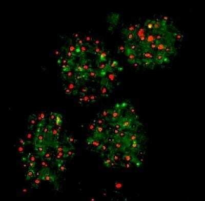
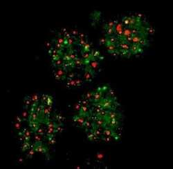
- Immunocytochemistry analysis of FANCD2 in SiHa cells after cell exposure to IR. Samples were incubated in FANCD2 polyclonal antibody (Product # PA1-16548). Analysis using the Biotin conjugate of FANCD2 Antibody. FANCD2 colocalizes in vivo with another protein in SiHa cells after cell exposure to IR. Proliferating SiHa cells were exposed to 10 Gy of IR and double color immunofluorescence staining was performed after 8 hours. Images were captured in a Kodak digital image system on a Leica fluorescence microscope.
- Submitted by
- Invitrogen Antibodies (provider)
- Main image
- Experimental details
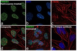
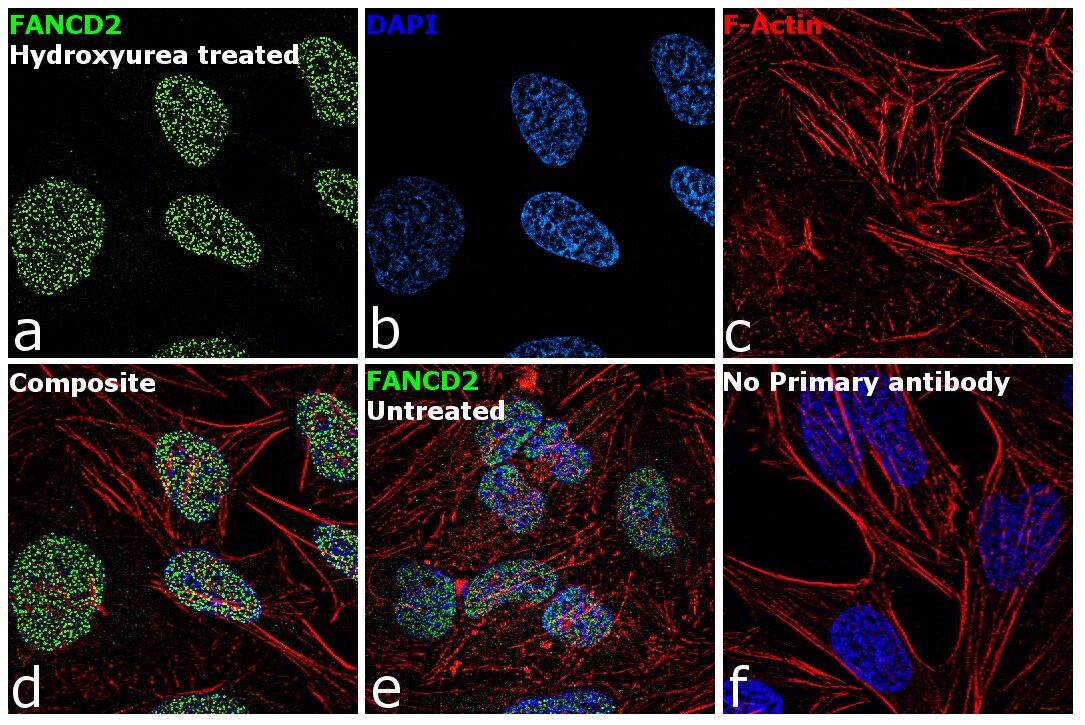
- Immunofluorescence analysis of FANCD2 was performed using 70% confluent log phase HeLa cells treated with 2mM of Hydroxyurea for 24 hours. The cells were fixed with 4% paraformaldehyde for 10 minutes, permeabilized with 0.1% Triton™ X-100 for 15 minutes, and blocked with 1% BSA for 1 hour at room temperature. The cells were labeled with FANCD2 Rabbit Polyclonal Antibody (Product # PA1-16548) at 5 µg/mL in 0.1% BSA, incubated at 4 degree Celsius overnight and then labeled with Goat anti-Rabbit IgG (H+L) Superclonal™ Secondary Antibody, Alexa Fluor® 488 conjugate (Product # A27034) at a dilution of 1:2000 for 45 minutes at room temperature (Panel a: green). Nuclei (Panel b: blue) were stained with ProLong™ Diamond Antifade Mountant with DAPI (Product # P36962). F-actin (Panel c: red) was stained with Rhodamine Phalloidin (Product # R415, 1:300). Panel d represents the merged image showing nuclear localization. Panel e shows untreated cells with reduced signal. Panel f represents control cells with no primary antibody to assess background. The images were captured at 60X magnification.
- Submitted by
- Invitrogen Antibodies (provider)
- Main image

- Experimental details
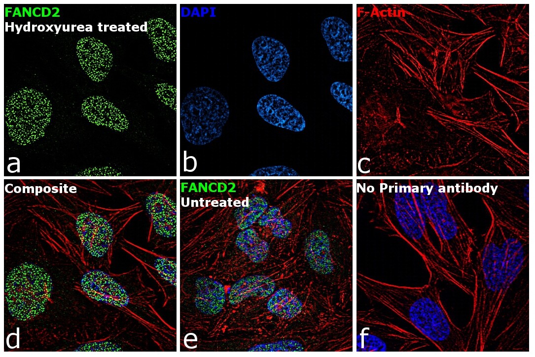
- Immunofluorescence analysis of FANCD2 was performed using 70% confluent log phase HeLa cells treated with 2mM of Hydroxyurea for 24 hours. The cells were fixed with 4% paraformaldehyde for 10 minutes, permeabilized with 0.1% Triton™ X-100 for 15 minutes, and blocked with 1% BSA for 1 hour at room temperature. The cells were labeled with FANCD2 Rabbit Polyclonal Antibody (Product # PA1-16548) at 5 µg/mL in 0.1% BSA, incubated at 4 degree Celsius overnight and then labeled with Goat anti-Rabbit IgG (Heavy Chain) Superclonal™ Secondary Antibody, Alexa Fluor® 488 conjugate (Product # A27034) at a dilution of 1:2000 for 45 minutes at room temperature (Panel a: green). Nuclei (Panel b: blue) were stained with ProLong™ Diamond Antifade Mountant with DAPI (Product # P36962). F-actin (Panel c: red) was stained with Rhodamine Phalloidin (Product # R415, 1:300). Panel d represents the merged image showing nuclear localization. Panel e shows untreated cells with reduced signal. Panel f represents control cells with no primary antibody to assess background. The images were captured at 60X magnification.
- Submitted by
- Invitrogen Antibodies (provider)
- Main image
- Experimental details
- Immunocytochemistry analysis of FANCD2 in SiHa cells after cell exposure to IR. Samples were incubated in FANCD2 polyclonal antibody (Product # PA1-16548). Analysis using the Biotin conjugate of FANCD2 Antibody. FANCD2 colocalizes in vivo with another protein in SiHa cells after cell exposure to IR. Proliferating SiHa cells were exposed to 10 Gy of IR and double color immunofluorescence staining was performed after 8 hours. Images were captured in a Kodak digital image system on a Leica fluorescence microscope.

Supportive validation
- Submitted by
- Invitrogen Antibodies (provider)
- Main image
- Experimental details
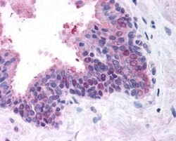
- Immunohistochemical analysis of FANCD2 in human prostate, glandular epithelium. Samples were incubated in FANCD2 polyclonal antibody (Product # PA1-16548). Image using the Biotin format of this antibody.

Supportive validation
- Submitted by
- Invitrogen Antibodies (provider)
- Main image
- Experimental details
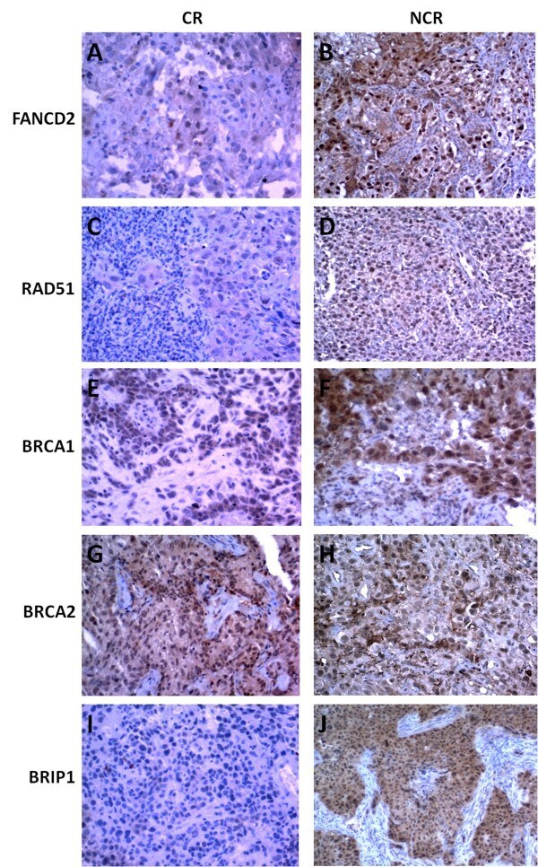
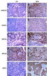
- Figure 5 Validation of FANCD2 (A-B), RAD51 (C-D), BRCA1 (E-F), BRCA2 (G-H) and BRIP1 (I-J) protein expression in advanced squamous cervical tumor cells. Staining for FANCD2, RAD51, BRCA1 and BRIP1 for the NCR cervical samples indicated strongly positive protein expression compared with the CR cervical samples. The BRCA2 protein expression was comparable between the NCR and CR cervical samples; (x200 magnification).