Explore
 Validate
 Learn
Learn Western blot
 Immunoprecipitation
Antibody data
- Antibody Data
- Antigen structure
- References [6]
- Comments [0]
- Validations
- Western blot [3]
- Immunocytochemistry [2]
- Immunohistochemistry [4]
Submit
Validation data
Reference
Comment
Report error
- Product number
- GTX101087 - Provider product page

- Provider
- GeneTex
- Proper citation
- GeneTex Cat#GTX101087, RRID:AB_1952549
- Product name
- Von Hippel Lindau antibody
- Antibody type
- Polyclonal
- Reactivity
- Human, Mouse, Rat
- Host
- Rabbit
Submitted references CXCR7 promotes melanoma tumorigenesis via Src kinase signaling.
The Breast Cancer Tumor Suppressor TRIM29 Is Expressed via ATM-dependent Signaling in Response to Hypoxia.
An ID2-dependent mechanism for VHL inactivation in cancer.
Minocycline accelerates hypoxia-inducible factor-1 alpha degradation and inhibits hypoxia-induced neovasculogenesis through prolyl hydroxylase, von Hippel-Lindau-dependent pathway.
The HILDA complex coordinates a conditional switch in the 3'-untranslated region of the VEGFA mRNA.
Silibinin inhibits VEGF secretion and age-related macular degeneration in a hypoxia-dependent manner through the PI-3 kinase/Akt/mTOR pathway.
Xu S, Tang J, Wang C, Liu J, Fu Y, Luo Y
Cell death & disease 2019 Feb 25;10(3):191
Cell death & disease 2019 Feb 25;10(3):191
The Breast Cancer Tumor Suppressor TRIM29 Is Expressed via ATM-dependent Signaling in Response to Hypoxia.
Dükel M, Streitfeld WS, Tang TC, Backman LR, Ai L, May WS, Brown KD
The Journal of biological chemistry 2016 Oct 7;291(41):21541-21552
The Journal of biological chemistry 2016 Oct 7;291(41):21541-21552
An ID2-dependent mechanism for VHL inactivation in cancer.
Lee SB, Frattini V, Bansal M, Castano AM, Sherman D, Hutchinson K, Bruce JN, Califano A, Liu G, Cardozo T, Iavarone A, Lasorella A
Nature 2016 Jan 14;529(7585):172-7
Nature 2016 Jan 14;529(7585):172-7
Minocycline accelerates hypoxia-inducible factor-1 alpha degradation and inhibits hypoxia-induced neovasculogenesis through prolyl hydroxylase, von Hippel-Lindau-dependent pathway.
Li CH, Liao PL, Yang YT, Huang SH, Lin CH, Cheng YW, Kang JJ
Archives of toxicology 2014 Mar;88(3):659-71
Archives of toxicology 2014 Mar;88(3):659-71
The HILDA complex coordinates a conditional switch in the 3'-untranslated region of the VEGFA mRNA.
Yao P, Potdar AA, Ray PS, Eswarappa SM, Flagg AC, Willard B, Fox PL
PLoS biology 2013;11(8):e1001635
PLoS biology 2013;11(8):e1001635
Silibinin inhibits VEGF secretion and age-related macular degeneration in a hypoxia-dependent manner through the PI-3 kinase/Akt/mTOR pathway.
Lin CH, Li CH, Liao PL, Tse LS, Huang WK, Cheng HW, Cheng YW
British journal of pharmacology 2013 Feb;168(4):920-31
British journal of pharmacology 2013 Feb;168(4):920-31
No comments: Submit comment
Supportive validation
- Submitted by
- GeneTex (provider)
- Main image
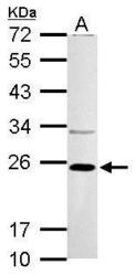
- Experimental details
- VHL antibody detects VHL protein by western blot analysis.A. 30 ?g Neuro2A whole cell lysate/extract B. 30 ?g GL261 whole cell lysate/extract 12% SDS-PAGEVHL antibody (GTX101087) dilution: 1:1000 The HRP-conjugated anti-rabbit IgG antibody (GTX213110-01) was used to detect the primary antibody.

- Submitted by
- GeneTex (provider)
- Main image
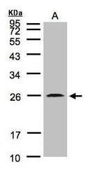
- Experimental details
- Sample(30 ?g of whole cell lysate)A:MOLT4(GTX27912)12% SDS PAGEGTX101087 diluted at 1:2000The HRP-conjugated anti-rabbit IgG antibody (GTX213110-01) was used to detect the primary antibody.
- Submitted by
- GeneTex (provider)
- Main image
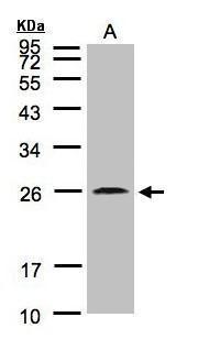
- Experimental details
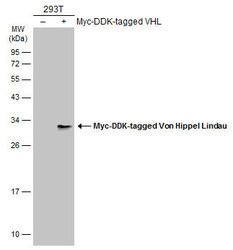
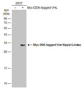
- Non-transfected (¡V) and transfected (+) 293T whole cell extracts (30 ?g) were separated by 12% SDS-PAGE, and the membrane was blotted with Von Hippel Lindau antibody (GTX101087) diluted at 1:5000. The HRP-conjugated anti-rabbit IgG antibody (GTX213110-01) was used to detect the primary antibody.
Supportive validation
- Submitted by
- GeneTex (provider)
- Main image
- Experimental details
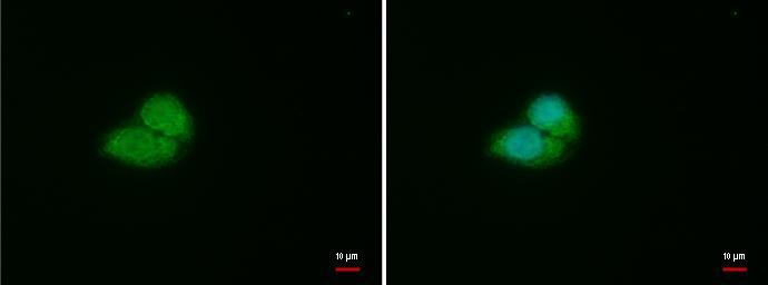
- VHL antibody detects VHL protein at cytoplasm and nucleus by immunofluorescent analysis.Sample: HepG2 cells were fixed in 4% paraformaldehyde at RT for 15 min.Green: VHL protein stained by VHL antibody (GTX101087) diluted at 1:500.Blue: Hoechst 33342 staining.
- Submitted by
- GeneTex (provider)
- Main image
- Experimental details
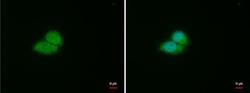
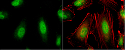
- VHL antibody detects VHL protein at cytoplasm and nucleus by immunofluorescent analysis.Sample: HeLa cells were fixed in 4% paraformaldehyde at RT for 15 min.Green: VHL protein stained by VHL antibody (GTX101087) diluted at 1:500.Red: phalloidin, a cytoskeleton marker, stained by phalloidin (invitrogen, A12380) diluted at 1:200.

Supportive validation
- Submitted by
- GeneTex (provider)
- Main image
- Experimental details
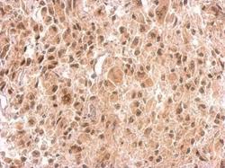
- VHL antibody detects VHL protein at on U373 xenograft by immunohistochemical analysis. Sample: Paraffin-embedded U373 xenograft. VHL antibody (GTX101087) dilution: 1:500.

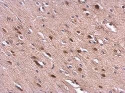
- Submitted by
- GeneTex (provider)
- Main image
- Experimental details
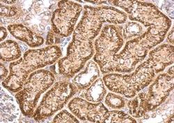
- VHL antibody detects VHL protein at cytosol on mouse kidney by immunohistochemical analysis. Sample: Paraffin-embedded mouse kidney. VHL antibody (GTX101087) dilution: 1:500.
- Submitted by
- GeneTex (provider)
- Main image
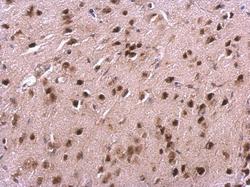
- Experimental details
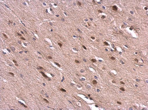
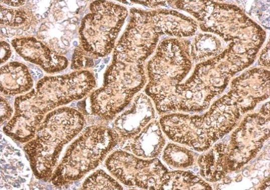
- VHL antibody detects VHL protein at cytosol and nucleus on mouse fore brain by immunohistochemical analysis. Sample: Paraffin-embedded mouse fore brain. VHL antibody (GTX101087) dilution: 1:500.
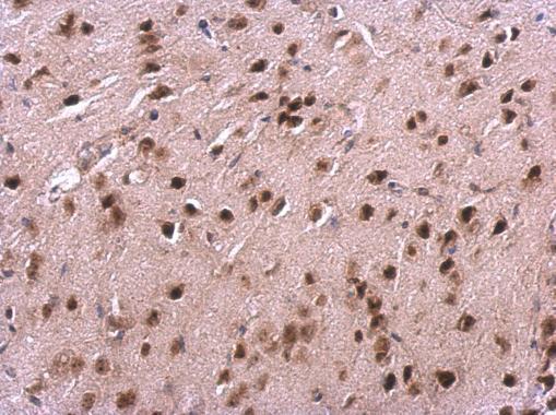
- Submitted by
- GeneTex (provider)
- Main image
- Experimental details
- VHL antibody detects VHL protein at cytosol and nucleus on rat fore brain by immunohistochemical analysis. Sample: Paraffin-embedded rat fore brain. VHL antibody (GTX101087) dilution: 1:500.