 Explore
 Validate
 Learn
Learn Western blot
 Immunohistochemistry
Antibody data
- Antibody Data
- Antigen structure
- References [0]
- Comments [0]
- Validations
- Western blot [3]
- Immunocytochemistry [1]
Submit
Validation data
Reference
Comment
Report error
- Product number
- MA1-90008 - Provider product page

- Provider
- Invitrogen Antibodies
- Product name
- PGP9.5 Monoclonal Antibody (13C4)
- Antibody type
- Monoclonal
- Antigen
- Other
- Description
- Recommended positive controls: Human Brain Tissue. It is recommended to perform heat mediated antigen retrieval before starting with the IHC protocol.
- Reactivity
- Human, Mouse, Rat
- Host
- Mouse
- Isotype
- IgG
- Antibody clone number
- 13C4
- Vial size
- 500 µL
- Concentration
- 0.3 mg/mL
- Storage
- 4° C, do not freeze
No comments: Submit comment
Supportive validation
- Submitted by
- Invitrogen Antibodies (provider)
- Main image
- Experimental details
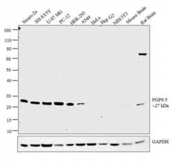
- Western blot analysis was performed on whole cell extracts (30 µg lysate) of Neuro-2a (Lane 1), SH-SY5Y (Lane 2), U-87 MG (Lane 3), PC-12 (Lane 4), HEK-293 (Lane 5), A549 (Lane 6), HeLa (Lane 7), Hep G2 (Lane 8), NIH/3T3 (Lane 9), tissue extracts of Mouse Brain (Lane 10) and Rat Brain (Lane 11). The blot was probed with Anti-PGP9.5 Monoclonal Antibody (Product # MA1-90008, 1:500 dilution) and detected by chemiluminescence using Goat anti-Mouse IgG (H+L) Superclonal™ Secondary Antibody, HRP conjugate (Product # A28177, 0.25 µg/ml, 1:4000 dilution). A 27 kDa band corresponding to PGP9.5 was detected across the cell lines and tissues tested except for Hela, HepG2 and NIH/3T3 which is reported to be negative for PGP9.5 expression.
- Submitted by
- Invitrogen Antibodies (provider)
- Main image

- Experimental details
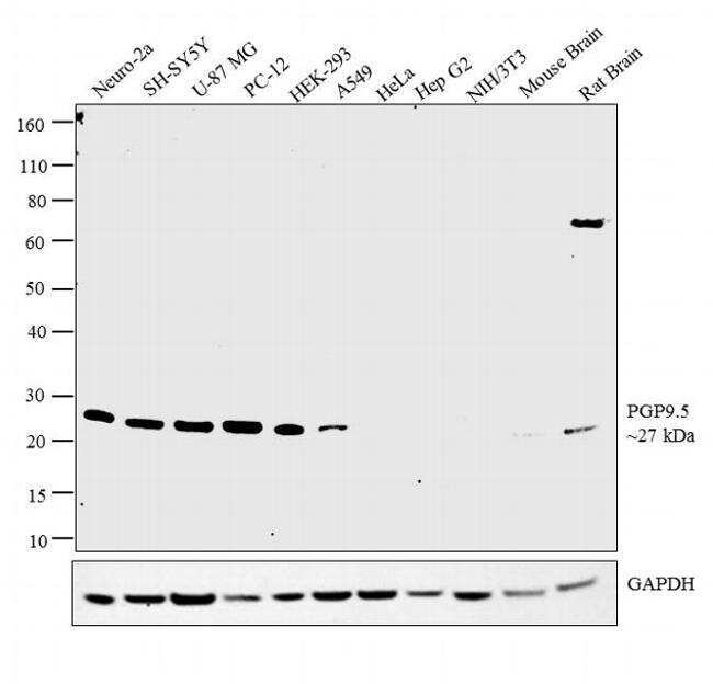
- Western blot analysis was performed on whole cell extracts (30 µg lysate) of Neuro-2a (Lane 1), SH-SY5Y (Lane 2), U-87 MG (Lane 3), PC-12 (Lane 4), HEK-293 (Lane 5), A549 (Lane 6), HeLa (Lane 7), Hep G2 (Lane 8), NIH/3T3 (Lane 9), tissue extracts of Mouse Brain (Lane 10) and Rat Brain (Lane 11). The blot was probed with Anti-PGP9.5 Monoclonal Antibody (Product # MA1-90008, 1:500 dilution) and detected by chemiluminescence using Goat anti-Mouse IgG (H+L) Superclonal™ Secondary Antibody, HRP conjugate (Product # A28177, 0.25 µg/ml, 1:4000 dilution). A 27 kDa band corresponding to PGP9.5 was detected across the cell lines and tissues tested except for Hela, HepG2 and NIH/3T3 which is reported to be negative for PGP9.5 expression.

- Submitted by
- Invitrogen Antibodies (provider)
- Main image

- Experimental details
- Western blot analysis of PGP9.5/UCHL1 was performed by loading 20 µg of SH-SY5Y wild type (Lane 1), SH-SY5Y Cas9 control (Lane 2), SH-SY5Y PGP9.5/UCHL1 knockout (Lane 3) whole cell extracts. The blot was probed with Anti-PGP9.5/ UCHL1 Monoclonal Antibody (Product # MA1-90008) (1:500 dilution) and Goat anti-Mouse IgG (H+L), Superclonal™ Recombinant Secondary Antibody, HRP (Product # A28177) (1:4000 dilution). Loss of signal upon CRISPR mediated knockout (KO) confirms that antibody is specific to PGP9.5/UCHL1.

Supportive validation
- Submitted by
- Invitrogen Antibodies (provider)
- Main image

- Experimental details
- Immunofluorescence analysis of PGP9.5 was performed using 70% confluent log phase SH-SY5Y cells. The cells were fixed with 4% paraformaldehyde for 10 minutes, permeabilized with 0.1% Triton™ X-100 for 10 minutes, and blocked with 1% BSA for 1 hour at room temperature. The cells were labeled with PGP9.5 Monoclonal Antibody (13C4) (Product # MA1-90008) at 5 µg/mL in 0.1% BSA and incubated overnight at 4 degree and then labeled with Goat anti-Mouse IgG (H+L) Superclonal™ Secondary Antibody, Alexa Fluor® 488 conjugate (Product # A28175) at a dilution of 1:2000 for 45 minutes at room temperature (Panel a: green). Nuclei (Panel b: blue) were stained with SlowFade® Gold Antifade Mountant with DAPI (Product # S36938). F-actin (Panel c: red) was stained with Rhodamine Phalloidin (Product # R415, 1:300). Panel d represents the merged image showing cytoplasmic localization. Panel e represents control cells with no primary antibody to assess background. The images were captured at 60X magnification.
