 Explore
 Validate
 Learn
Learn Western blot
 Immunohistochemistry
Antibody data
- Antibody Data
- Antigen structure
- References [0]
- Comments [0]
- Validations
- Western blot [4]
- Immunocytochemistry [1]
Submit
Validation data
Reference
Comment
Report error
- Product number
- PA1-10024 - Provider product page

- Provider
- Invitrogen Antibodies
- Product name
- PGP9.5 Polyclonal Antibody
- Antibody type
- Polyclonal
- Antigen
- Purifed from natural sources
- Description
- PA1-10024 was made against full length recombinant human UCHL1 expressed in and purified from E. coli and can be used to identify neurons and their processes in culture or in sections. The antibody works cleanly on appropriate lysates of cell and tissues.
- Reactivity
- Human, Mouse, Rat, Bovine, Chicken/Avian, Porcine
- Host
- Rabbit
- Isotype
- IgG
- Vial size
- 100 µL
- Concentration
- Conc. Not Determined
- Storage
- Store at 4°C short term. For long term storage, store at -20°C, avoiding freeze/thaw cycles.
No comments: Submit comment
Supportive validation
- Submitted by
- Invitrogen Antibodies (provider)
- Main image

- Experimental details
- Western blot was performed using Anti-PGP9.5 Polyclonal Antibody, (Product # PA1-10024) and a ~25 kDa band corresponding to PGP9.5 was observed across the panel tested except SW480, HT-29, HeLa and HepG2 which are reported to be negative. Whole cell extracts (30 µg lysate) of SH-SY5Y (Lane 1), U-87 MG (Lane 2), IMR-32 (Lane 3), SK-N-SH (Lane 4), A549 (Lane 5), SW480 (Lane 6), HT-29 (Lane 7), HeLa (Lane 8), HepG2 (Lane 9) and Neuro-2a (Lane 10) were electrophoresed using Novex® NuPAGE® 12% % Bis-Tris gel (Product # NP0341BOX). Resolved proteins were then transferred onto a nitrocellulose membrane (Product # IB23001) by iBlot® 2 Dry Blotting System (Product # IB21001). The blot was probed with the primary antibody (1:2000 dilution) and detected by chemiluminescence with Goat anti-Rabbit IgG (H+L) Superclonal™ Recombinant Secondary Antibody, HRP (Product # A27036, 1:4000 dilution) using the iBright FL 1000 (Product # A32752). Chemiluminescent detection was performed using Novex® ECL Chemiluminescent Substrate Reagent Kit (Product # WP20005).

- Submitted by
- Invitrogen Antibodies (provider)
- Main image
- Experimental details
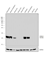
- Western blot was performed using Anti-PGP9.5 Polyclonal antibody, (Product # PA1-10024) and a ~25 kDa band corresponding to PGP9.5 was observed across the Mouse and Rat tissues except Mouse and Rat Liver and Heart which are reported to be negative. Tissue extracts (30 µg lysate) of Mouse Brain (Lane1), Mouse Cerebellum (Lane 2), Mouse Eye (Lane 3), Mouse Liver (Lane 4), Mouse Heart (Lane 5), Rat Brain (Lane 6), Rat Cerebellum (Lane 7), Rat Liver (Lane 8) and Rat Heart (Lane 9) were electrophoresed using Novex® NuPAGE® 12 % Bis-Tris gel (Product # NP0341BOX). Resolved proteins were then transferred onto a nitrocellulose membrane (Product # IB23001) by iBlot® 2 Dry Blotting System (Product # IB21001). The blot was probed with the primary antibody (1:2000 dilution) and detected by chemiluminescence with Goat anti-Rabbit IgG (H+L) Superclonal™ Recombinant Secondary Antibody, HRP (Product # A27036, 1:4000 dilution) using the iBright FL 1000 (Product # A32752). Chemiluminescent detection was performed using Novex® ECL Chemiluminescent Substrate Reagent Kit (Product # WP20005).

- Submitted by
- Invitrogen Antibodies (provider)
- Main image

- Experimental details
- Western blot analysis of PGP9.5 in different tissue and cell lysates using UCHL1 polyclonal antibody (Product # PA1-10024) at a dilution of 1:2,000 as seen in red, and an HSP60 monoclonal antibody at a dilution of 1:10,000 as seen in green. 1) protein standard, 2) rat brain, 3) mouse brain, 4) NIH-3T3, 5) HEK293, 6) HeLa, 7) SH-SY5Y cells. The single band at 24kDa corresponds to the UCHL1 protein, while the 60kDa band represents HSP60 protein. UCHL1 is detectable in CNS extracts and cells with neuronal properties but not in HeLa, NIH-3T3 and other non-neuronal cells.

- Submitted by
- Invitrogen Antibodies (provider)
- Main image
- Experimental details
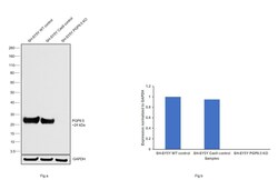
- Western blot analysis of PGP9.5/UCHL1 was performed by loading 20 µg of SH-SY5Y wild type (Lane 1), SH-SY5Y Cas9 control (Lane 2), SH-SY5Y PGP9.5/UCHL1 knockout (Lane 3) whole cell extracts. The blot was probed with Anti-PGP9.5/ UCHL1 Polyclonal Antibody (Product # PA1-10024) (1:5000 dilution) and Goat anti-Rabbit IgG (H+L), Superclonal™ Recombinant Secondary Antibody, HRP (Product # A27036) (1:4000 dilution). Loss of signal upon CRISPR mediated knockout (KO) confirms that antibody is specific to PGP9.5/UCHL1.

Supportive validation
- Submitted by
- Invitrogen Antibodies (provider)
- Main image

- Experimental details
- Immunofluorescent analysis of PGP9.5 in SH-SY5Y cells. The cells were stained using a UCHL1 polyclonal antibody (Product # PA1-10024) at a dilution of 1:1,000 as seen in green, with a fibrillarin monoclonal antibody at a dilution of 1:1,000 as seen in red, and with DAPI staining the nuclear DNA in blue. The UCHL1 antibody produces strong staining of the cellular cytoplasm of these cells which share many properties with neurons, while the fibrillarin antibody specifically labels nucleoli.
