 Explore
 Validate
 Learn
Learn Western blot
 ELISA
ELISAAntibody data
- Antibody Data
- Antigen structure
- References [0]
- Comments [0]
- Validations
- Western blot [4]
- Immunohistochemistry [1]
- Flow cytometry [1]
Submit
Validation data
Reference
Comment
Report error
- Product number
- NBP2-33130 - Provider product page

- Provider
- Novus Biologicals
- Product name
- Mouse Monoclonal UCH-L1/PGP9.5 Antibody
- Antibody type
- Monoclonal
- Description
- Protein G purified. This MAb reacts with a protein of 20-30kDa, identified as PGP9.5, also known as ubiquitin carboxyl-terminal hydrolase-1 (UchL1). Initially, PGP9.5 expression in normal tissues was reported in neurons and neuroendocrine cells but later it was found in distal renal tubular epithelium, spermatogonia, Leydig cells, oocytes, melanocytes, prostatic secretory epithelium, ejaculatory duct cells, epididymis, mammary epithelial cells, Merkel cells, and dermal fibroblasts. Furthermore, immunostaining for PGP9.5 has been shown in a wide variety of mesenchymal neoplasms as well. A mutation in PGP9.5 gene is believed to cause a form of Parkinson's disease.
- Reactivity
- Human, Mouse, Rat, Bovine, Porcine, Zebrafish
- Host
- Mouse
- Isotype
- IgG
- Vial size
- 0.1 mg
- Concentration
- 1.0 mg/ml
- Storage
- Store at 4C short term. Aliquot and store at -20C long term. Avoid freeze-thaw cycles.
No comments: Submit comment
Supportive validation
- Submitted by
- Novus Biologicals (provider)
- Main image

- Experimental details
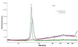
- Simple Western: UCH-L1/PGP9.5 Antibody (31A3) - Azide and BSA Free [NBP2-33130] - Simple Western lane view shows a specific band for PGP9.5 / UCHL-1 in 0.2 mg/ml of h. Cerebellum (left) and IMR-32 (right) lysate(s). This experiment was performed under reducing conditions using the 12-230 kDa separation system.

- Submitted by
- Novus Biologicals (provider)
- Main image
- Experimental details
- Simple Western: UCH-L1/PGP9.5 Antibody (31A3) - Azide and BSA Free [NBP2-33130] - Electropherogram images of the corresponding Simple Western lane. PGP9.5 / UCHL-1 antibody was used at 10 ug/ml dilution of h. Cerebellum and IMR-32 lysates(s) respectively.

- Submitted by
- Novus Biologicals (provider)
- Main image
- Experimental details
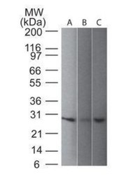
- Western Blot: UCH-L1/PGP9.5 Antibody (31A3) - Azide and BSA Free [NBP2-33130] - analysis of UchL1 in 1) human, 2) mouse and 3) rat brain lysate using UchL1 antibody at 1 ug/ml. goat anti-mouse Ig HRP secondary antibody and ECL substrate solution were used for this test.

- Submitted by
- Novus Biologicals (provider)
- Main image
- Experimental details
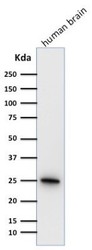
- Western Blot: UCH-L1/PGP9.5 Antibody (31A3) - Azide and BSA Free [NBP2-33130] - Western Blot Analysis of human brain tissue lysate using UCH-L1/PGP9.5 Antibody (31A3)

Supportive validation
- Submitted by
- Novus Biologicals (provider)
- Main image

- Experimental details
- Immunohistochemistry-Paraffin: UCH-L1/PGP9.5 Antibody (31A3) - Azide and BSA Free [NBP2-33130] - Formalin-fixed, paraffin-embedded human brain stained with UchL1 antibody (PGP9.5) (1:500), peroxidase-conjugate and DAB chromogen. Staining seen in cytoplasm, ER and membrane. Fixation in 95% ethanol/5% acetic acid for 2-3 hours prior to paraffin embedd

Supportive validation
- Submitted by
- Novus Biologicals (provider)
- Main image

- Experimental details
- Flow Cytometry: UCH-L1/PGP9.5 Antibody (31A3) - Azide and BSA Free [NBP2-33130] - Flow Cytometric Analysis of T98G cells using UCH-L1/PGP9.5 Antibody (31A3)followed by Goat anti-Mouse IgG-CF488 (Blue); Isotype Control (Red).
