 Explore
 Validate
 Learn
Learn Western blot
Antibody data
- Antibody Data
- Antigen structure
- References [0]
- Comments [0]
- Validations
- Western blot [7]
- Immunocytochemistry [3]
- Immunohistochemistry [1]
Submit
Validation data
Reference
Comment
Report error
- Product number
- MA1-46079 - Provider product page

- Provider
- Invitrogen Antibodies
- Product name
- PGP9.5 Monoclonal Antibody (BH7)
- Antibody type
- Monoclonal
- Antigen
- Purifed from natural sources
- Description
- Suggested positive control: bovine brain whole cell homogenate, SHSY-5Y or HEK 293 cell extract, and rat spinal cord/peripheral nerve homogenate, antigen standard for UCHL1 (transient overexpression lysate).
- Reactivity
- Human, Mouse, Rat, Bovine, Porcine
- Host
- Mouse
- Isotype
- IgG
- Antibody clone number
- BH7
- Vial size
- 100 µL
- Concentration
- 1 mg/mL
- Storage
- Store at 4°C short term. For long term storage, store at -20°C, avoiding freeze/thaw cycles.
No comments: Submit comment
Supportive validation
- Submitted by
- Invitrogen Antibodies (provider)
- Main image

- Experimental details
- Western blot of whole cell homogenate of bovine brain stained with MA1-46079 (right most lane) and two other monoclonal antibodies reactive with UCHL1 (left and central lane). All three antibodies show a single clean band running at about 24kDa.

- Submitted by
- Invitrogen Antibodies (provider)
- Main image

- Experimental details
- Western blot analysis of PGP9.5 in tissue lysates. Samples were incubated in PGP9.5 monoclonal antibody (Product # MA1-46079 using a dilution of 1:10,000. Antibody in green: [1] protein standard (red), [2] rat brain, [3] rat spinal cord, [4] mouse brain, [5] mouse spinal cord, [6] pig brain, [7] pig spinal cord. The single band at 24 kDa corresponds to the UCHL1 protein.

- Submitted by
- Invitrogen Antibodies (provider)
- Main image
- Experimental details
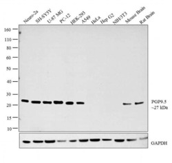
- Western blot analysis was performed on whole cell extracts (30 µg lysate) of Neuro-2a (Lane 1), SH-SY5Y (Lane 2), U-87 MG (Lane 3), PC-12 (Lane 4), HEK-293 (Lane 5), A549 (Lane 6), HeLa (Lane 7), Hep G2 (Lane 8), NIH/3T3 (Lane 9), tissue extracts of Mouse Brain (Lane 10) and Rat Brain (Lane 11). The blot was probed with Anti-PGP9.5 Monoclonal Antibody (Product # MA1-46079, 1:10,000 dilution) and detected by chemiluminescence using Goat anti-Mouse IgG (H+L) Superclonal™ Secondary Antibody, HRP conjugate (Product # A28177, 0.25 µg/ml, 1:4000 dilution). A 27 kDa band corresponding to PGP9.5 was detected across the cell lines and tissues tested except for Hela, HepG2 and NIH/3T3 which is reported to be negative for PGP9.5 expression.

- Submitted by
- Invitrogen Antibodies (provider)
- Main image

- Experimental details
- Western blot analysis of PGP9.5 in tissue lysates. Samples were incubated in PGP9.5 monoclonal antibody (Product # MA1-46079 using a dilution of 1:10,000. Antibody in green: [1] protein standard (red), [2] rat brain, [3] rat spinal cord, [4] mouse brain, [5] mouse spinal cord, [6] pig brain, [7] pig spinal cord. The single band at 24 kDa corresponds to the UCHL1 protein.

- Submitted by
- Invitrogen Antibodies (provider)
- Main image

- Experimental details
- Western blot analysis of PGP9.5 in 0.02 mg/mL Human Brain lysate. Samples were incubated in PGP9.5 monoclonal antibody (Product # MA1-46079). This experiment was performed under reducing conditions using the 12-230 kDa separation system.

- Submitted by
- Invitrogen Antibodies (provider)
- Main image

- Experimental details
- Western blot analysis of PGP9.5/UCHL1 was performed by loading 20 µg of SH-SY5Y wild type (Lane 1), SH-SY5Y Cas9 control (Lane 2), SH-SY5Y PGP9.5/UCHL1 knockout (Lane 3) whole cell extracts. The blot was probed with Anti-PGP9.5/ UCHL1 Monoclonal Antibody (Product # MA1-46079) (1:10000 dilution) and Goat anti-Mouse IgG (H+L), Superclonal™ Recombinant Secondary Antibody, HRP (Product # A28177) (1:4000 dilution). Loss of signal upon CRISPR mediated knockout (KO) confirms that antibody is specific to PGP9.5/UCHL1.

Supportive validation
- Submitted by
- Invitrogen Antibodies (provider)
- Main image

- Experimental details
- Immunocytochemistry analysis of PGP9.5 in human embryonic kidney cell line 293. Samples were incubated in PGP9.5 monoclonal antibody (Product # MA1-46079). This antibody (green) and rabbit antibody to neurofilament NF-M (red). Blue is a DNA stain. Note that some of the Hek293 cells, which perhaps surprisingly have some properties of neuronal lineage cells (4), are beginning to express both UCHL1 and NF-M, both markers of neurons. The majority of cells, however express neither protein. Note that the UCHL1 is cytoplasmic and diffuse while the NF-M is concentrated in cytoplasmic filaments.

- Submitted by
- Invitrogen Antibodies (provider)
- Main image
- Experimental details
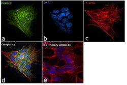
- Immunofluorescence analysis of PGP9.5 was performed using 70% confluent log phase SH-SY5Y cells. The cells were fixed with 4% paraformaldehyde for 10 minutes, permeabilized with 0.1% Triton™ X-100 for 10 minutes, and blocked with 1% BSA for 1 hour at room temperature. The cells were labeled with PGP9.5 Monoclonal Antibody (BH7) (Product # MA1-46079) at 1:1000 dilution in 0.1% BSA and incubated overnight at 4 degree and then labeled with Goat anti-Mouse IgG (H+L) Superclonal™ Secondary Antibody, Alexa Fluor® 488 conjugate (Product # A28175) at a dilution of 1:2000 for 45 minutes at room temperature (Panel a: green). Nuclei (Panel b: blue) were stained with SlowFade® Gold Antifade Mountant with DAPI (Product # S36938). F-actin (Panel c: red) was stained with Rhodamine Phalloidin (Product # R415, 1:300). Panel d represents the merged image showing and cytoplasmic localization. Panel e represents control cells with no primary antibody to assess background. The images were captured at 60X magnification.

Supportive validation
- Submitted by
- Invitrogen Antibodies (provider)
- Main image

- Experimental details
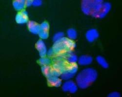
- Immunohistochemical analysis of PGP9.5 in rat hippocampal sections. Samples were incubated in PGP9.5 monoclonal antibody (Product # MA1-46079) using a dilution of 1:5,000 (green). Costained with rabbit pAb to FOX/NeuN, dilution 1:2,000, in red. The blue is DAPI staining of nuclear DNA. Following transcardial perfusion of rat with 4% paraformaldehyde, brain was post fixed for 24 hours, cut to 45 µM, and free-floating sections were stained with above antibodies. The UCHL1 antibody stains the cell body and dendrites of hippocampal neurons, while the FOX3 antibody labels nuclei of the neuronal cells.
