 Explore
 Validate
 Learn
Learn Western blot
Antibody data
- Antibody Data
- Antigen structure
- References [4]
- Comments [0]
- Validations
- Western blot [3]
- Immunocytochemistry [3]
- Other assay [1]
Submit
Validation data
Reference
Comment
Report error
- Product number
- PA1-10011 - Provider product page

- Provider
- Invitrogen Antibodies
- Product name
- PGP9.5 Polyclonal Antibody
- Antibody type
- Polyclonal
- Antigen
- Recombinant full-length protein
- Description
- PA1-10011 was made against full length recombinant human UCHL1 expressed in and purified from E. coli and can be used to identify neurons and their processes in culture or in sections. The antibody works cleanly on appropriate lysates of cell and tissues. |Stable at 4°C.
- Reactivity
- Human, Mouse, Rat, Bovine, Porcine
- Host
- Chicken/Avian
- Isotype
- IgY
- Vial size
- 100 µL
- Concentration
- Conc. Not Determined
- Storage
- 4° C
Submitted references Methods to Study the Myenteric Plexus of Rat Small Intestine.
Oxidative DNA Damage and Cisplatin Neurotoxicity Is Exacerbated by Inhibition of OGG1 Glycosylase Activity and APE1 Endonuclease Activity in Sensory Neurons.
Activation of GPR18 by Resolvin D2 Relieves Pain and Improves Bladder Function in Cyclophosphamide-Induced Cystitis Through Inhibiting TRPV1.
Acute Exposure to the Food-Borne Pathogen Listeria monocytogenes Does Not Induce α-Synuclein Pathology in the Colonic ENS of Nonhuman Primates.
Hecking I, Stegemann LN, Stahlke S, Theis V, Vorgerd M, Matschke V, Theiss C
Cellular and molecular neurobiology 2023 Jan;43(1):315-325
Cellular and molecular neurobiology 2023 Jan;43(1):315-325
Oxidative DNA Damage and Cisplatin Neurotoxicity Is Exacerbated by Inhibition of OGG1 Glycosylase Activity and APE1 Endonuclease Activity in Sensory Neurons.
Behrouzi A, Xia H, Thompson EL, Kelley MR, Fehrenbacher JC
International journal of molecular sciences 2022 Feb 8;23(3)
International journal of molecular sciences 2022 Feb 8;23(3)
Activation of GPR18 by Resolvin D2 Relieves Pain and Improves Bladder Function in Cyclophosphamide-Induced Cystitis Through Inhibiting TRPV1.
Lu Q, Yang Y, Zhang H, Chen C, Zhao J, Yang Z, Fan Y, Li L, Feng H, Zhu J, Yi S
Drug design, development and therapy 2021;15:4687-4699
Drug design, development and therapy 2021;15:4687-4699
Acute Exposure to the Food-Borne Pathogen Listeria monocytogenes Does Not Induce α-Synuclein Pathology in the Colonic ENS of Nonhuman Primates.
Mancinelli AM, Vichich JM, Zinnen AD, Hugon AM, Bondarenko V, Metzger JM, Simmons HA, Golos TG, Emborg ME
Journal of inflammation research 2021;14:7265-7279
Journal of inflammation research 2021;14:7265-7279
No comments: Submit comment
Supportive validation
- Submitted by
- Invitrogen Antibodies (provider)
- Main image

- Experimental details
- Western blot analysis was performed on whole cell extracts (30 µg lysate) of Neuro-2a (Lane 1), SH-SY5Y (Lane 2), U-87 MG (Lane 3), PC-12 (Lane 4), HEK-293 (Lane 5), A549 (Lane 6), HeLa (Lane 7), Hep G2 (Lane 8), NIH/3T3 (Lane 9), tissue extracts of Mouse Brain (Lane 10) and Rat Brain (Lane 11). The blot was probed with Anti-PGP9.5 Polyclonal Antibody (Product # PA1-10011, 1:10,000 dilution) and detected by chemiluminescence using Goat anti-Chicken IgY (H+L) Secondary Antibody, HRP (Product # A16054, 0.25 µg/ml, 1:4000 dilution). A 27 kDa band corresponding to PGP9.5 was detected across the cell lines and tissues tested except for Hela, HepG2 and NIH/3T3 which is reported to be negative for PGP9.5 expression.

- Submitted by
- Invitrogen Antibodies (provider)
- Main image

- Experimental details
- Western blot analysis of PGP9.5 in tissue and cell lysates using a PGP9.5 polyclonal antibody (Product # PA1-10011) at a dilution of 1:2,000 as seen in green, and using an Actin monoclonal antibody at a dilution of 1:1,000 as seen in red. 1) protein standard, 2) rat brain, 3) mouse brain, 4) NIH-3T3, 5) HEK293, 6) HeLa and 7) SH-SY5Y cells. The single band at 24 kDa mark corresponds to PGP9.5 protein which is detectable in CNS extracts and lysates of cells with neuronal properties but not in lysates of HeLa, NIH-3T3 and other non-neuronal cells. Actin is detected with apparent molecular weight of 42 kDa and provides an excellent loading control.

- Submitted by
- Invitrogen Antibodies (provider)
- Main image

- Experimental details
- Western blot analysis of PGP9.5/UCHL1 was performed by loading 20 µg of SH-SY5Y wild type (Lane 1), SH-SY5Y Cas9 control (Lane 2), SH-SY5Y PGP9.5/UCHL1 knockout (Lane 3) whole cell extracts. The blot was probed with Anti-PGP9.5/ UCHL1 Polyclonal Antibody (Product # PA1-10011) (1:10000 dilution) and Goat anti-Chicken IgY (H+L) Secondary Antibody, HRP (Product # A16054) (1:4000 dilution). Loss of signal upon CRISPR mediated knockout (KO) confirms that antibody is specific to PGP9.5/UCHL1.

Supportive validation
- Submitted by
- Invitrogen Antibodies (provider)
- Main image
- Experimental details
- Immunofluorescent analysis of PGP9.5 using a polyclonal antibody (Product # PA1-10011).

- Submitted by
- Invitrogen Antibodies (provider)
- Main image
- Experimental details
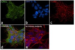
- Immunofluorescence analysis of PGP9.5 was performed using 70% confluent log phase SH-SY5Y cells. The cells were fixed with 4% paraformaldehyde for 10 minutes, permeabilized with 0.1% Triton™ X-100 for 10 minutes, and blocked with 1% BSA for 1 hour at room temperature. The cells were labeled with PGP9.5 Chicken Polyclonal Antibody (Product # PA1-10011) at 1:1000 dilution in 0.1% BSA and incubated overnight at 4 degree and then labeled with Goat anti-Chicken IgY (H+L) Secondary Antibody, Alexa Fluor 488 conjugate (Product # A28175) at a dilution of 1:2000 for 45 minutes at room temperature (Panel a: green). Nuclei (Panel b: blue) were stained with SlowFade® Gold Antifade Mountant with DAPI (Product # S36938). F-actin (Panel c: red) was stained with Rhodamine Phalloidin (Product # R415, 1:300). Panel d represents the merged image showing and cytoplasmic localization. Panel e represents control cells with no primary antibody to assess background. The images were captured at 60X magnification.

- Submitted by
- Invitrogen Antibodies (provider)
- Main image

- Experimental details
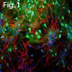
- Immunofluorescent analysis of PGP9.5 in cortical neuron-glial cell culture. The culture was prepared from an E20 rat and stained using a PGP9.5 polyclonal antibody (Product # PA1-10011) at a dilution of 1:500 as seen in red, and costained using a Vimentin monoclonal antibody at a dilution 1:2,000 as seen in green, and with DAPI staining the nuclear DNA in blue. The PGP9.5 antibody produces strong staining of the cell body and dendrites in neurons. The vimentin antibody stains intermediate filaments in fibroblastic and developing glial cells.

Supportive validation
- Submitted by
- Invitrogen Antibodies (provider)
- Main image

- Experimental details
- Figure 3 The expression of GPR18 was decreased in the bladders and DRGs of rats with CYP-induced cystitis 4, 24, and 48 h after intraperitoneal injection of saline or CYP. ( A ) Analysis of the mRNA expression of GPR18 in the bladders (n=6). ( B and C ) Analysis of the protein expression of GPR18 and representative Western blotting images of GPR18 in the bladder (n=6). ( D ) Analysis of the coexpression of GPR18 (red) and PGP9.5 (green) in normal bladder sections using immunofluorescence. ( E ) Analysis of the mRNA expression of GPR18 in the L6-S1 DRGs (n=6). ( F and G ) Analysis of the expression and distribution of GPR18 in the L6-S1 DRGs using immunofluorescence (n=6). The arrows indicate the afferent nerves that supply the bladder. Student's t -test was used for statistical analysis. *P
