 Explore
 Validate
 Learn
Learn Western blot
 Immunohistochemistry
Antibody data
- Antibody Data
- Antigen structure
- References [1]
- Comments [0]
- Validations
- Western blot [3]
- Immunocytochemistry [2]
Submit
Validation data
Reference
Comment
Report error
- Product number
- PA1-46204 - Provider product page

- Provider
- Invitrogen Antibodies
- Product name
- PGP9.5 Polyclonal Antibody
- Antibody type
- Polyclonal
- Antigen
- Purifed from natural sources
- Description
- Suggested positive control: antigen standard for UCHL1 (transient overexpression lysate), rat spinal cord or perpheral nerve homogenate.
- Reactivity
- Human, Mouse, Rat, Bovine, Porcine
- Host
- Chicken/Avian
- Isotype
- IgY
- Vial size
- 100 µL
- Concentration
- Conc. Not Determined
- Storage
- Store at 4°C short term. For long term storage, store at -20°C, avoiding freeze/thaw cycles.
Submitted references Genetic Mouse Models to Study Pancreatic Cancer-Induced Pain and Reduction in Well-Being.
Hirth M, Xie Y, Höper C, Prats A, Hackert T, Ebert MP, Kuner R
Cells 2022 Aug 24;11(17)
Cells 2022 Aug 24;11(17)
No comments: Submit comment
Supportive validation
- Submitted by
- Invitrogen Antibodies (provider)
- Main image
- Experimental details
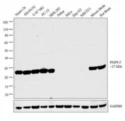
- Western blot analysis was performed on whole cell extracts (30 µg lysate) of Neuro-2a (Lane 1), SH-SY5Y (Lane 2), U-87 MG (Lane 3), PC-12 (Lane 4), HEK-293 (Lane 5), Jurkat (Lane 6), HeLa (Lane 7), Hep G2 (Lane 8), NIH/3T3 (Lane 9), tissue extracts of Mouse Brain (Lane 10) and Rat Brain (Lane 11). The blot was probed with Anti-PGP9.5 Polyclonal Antibody (Product # PA1-46204, 1:1000 dilution) and detected by chemiluminescence using Goat anti- Chicken IgG (H+L) Superclonal™ Secondary Antibody, HRP conjugate (Product # A16054, 0.25 µg/ml, 1:4000 dilution). A 27 kDa band corresponding to PGP9.5 was detected across the cell lines and tissues tested except for Jurkat, Hela, HepG2 and NIH/3T3 which is reported to be negative for PGP9.5 expression.

- Submitted by
- Invitrogen Antibodies (provider)
- Main image
- Experimental details
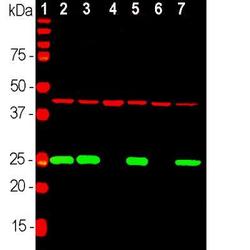
- Western blot analysis of PGP9.5 in rat brain, mouse brain, NIH-3T3, HEK293, HeLa and SH-SY5Y cells. Sample was incubated in PGP9.5 polyclonal antibody (Product # PA1-46204).

- Submitted by
- Invitrogen Antibodies (provider)
- Main image
- Experimental details
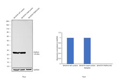
- Western blot analysis of PGP9.5/UCHL1 was performed by loading 20 µg of SH-SY5Y wild type (Lane 1), SH-SY5Y Cas9 control (Lane 2), SH-SY5Y PGP9.5/UCHL1 knockout (Lane 3) whole cell extracts. The blot was probed with Anti-PGP9.5/ UCHL1 Polyclonal Antibody (Product # PA1-46204) (1:5000 dilution) and Goat anti-Chicken IgY (H+L) Secondary Antibody, HRP (Product # A16054) (1:4000 dilution). Loss of signal upon CRISPR mediated knockout (KO) confirms that antibody is specific to PGP9.5/UCHL1.

Supportive validation
- Submitted by
- Invitrogen Antibodies (provider)
- Main image

- Experimental details
- Immunocytochemistry analysis of PGP9.5 in cortical neuron-glial cell culture from E20 rat. Samples were incubated in PGP9.5 polyclonal antibody (Product # PA1-46204) using a dilution of 1:500. This antibody in red, and costained with mouse mAb to vimentin, dilution 1:2,000, in green. The blue is DAPI staining of nuclear DNA. The UCHL1 antibody produces strong staining of the cell body and dendrites in neurons. The vimentin antibody stains intermediate filaments in fibroblastic and developing glial cells.

- Submitted by
- Invitrogen Antibodies (provider)
- Main image
- Experimental details
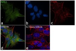
- Immunofluorescence analysis of PGP9.5 was performed using 70% confluent log phase SH-SY5Y cells. The cells were fixed with 4% paraformaldehyde for 10 minutes, permeabilized with 0.1% Triton™ X-100 for 10 minutes, and blocked with 1% BSA for 1 hour at room temperature. The cells were labeled with PGP9.5 Chicken Polyclonal Antibody (Product # PA1-46204) at 1:1000 dilution in 0.1% BSA and incubated overnight at 4 degree and then labeled with Goat anti-Chicken IgY (H+L) Secondary Antibody, Alexa Fluor 488 conjugate (Product # A10039) at a dilution of 1:2000 for 45 minutes at room temperature (Panel a: green). Nuclei (Panel b: blue) were stained with SlowFade® Gold Antifade Mountant with DAPI (Product # S36938). F-actin (Panel c: red) was stained with Rhodamine Phalloidin (Product # R415, 1:300). Panel d represents the merged image showing and cytoplasmic localization. Panel e represents control cells with no primary antibody to assess background. The images were captured at 60X magnification.
