 Explore
 Validate
 Learn
Learn Western blot
 Immunohistochemistry
Antibody data
- Antibody Data
- Antigen structure
- References [0]
- Comments [0]
- Validations
- Western blot [6]
- Immunocytochemistry [4]
Submit
Validation data
Reference
Comment
Report error
- Product number
- PA1-46205 - Provider product page

- Provider
- Invitrogen Antibodies
- Product name
- PGP9.5 Polyclonal Antibody
- Antibody type
- Polyclonal
- Antigen
- Purifed from natural sources
- Description
- Suggested positive control: antigen standard for UCHL1 (transient overexpression lysate), SH-SY5Y neuroblastoma lysate and rat spinal cord and peripheral nerve homogenate.
- Reactivity
- Human, Mouse, Rat, Bovine, Chicken/Avian, Porcine
- Host
- Rabbit
- Isotype
- IgG
- Vial size
- 100 µL
- Concentration
- Conc. Not Determined
- Storage
- Store at 4°C short term. For long term storage, store at -20°C, avoiding freeze/thaw cycles.
No comments: Submit comment
Supportive validation
- Submitted by
- Invitrogen Antibodies (provider)
- Main image

- Experimental details
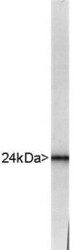
- Western blot of whole bovine brain extract stained with RPCA-UCHL1 showing a strong and clean band at about 24kDa.

- Submitted by
- Invitrogen Antibodies (provider)
- Main image
- Experimental details
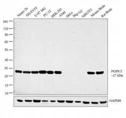
- Western blot analysis was performed on whole cell extracts (30 µg lysate) of Neuro-2a (Lane 1), SH-SY5Y (Lane 2), U-87 MG (Lane 3), PC-12 (Lane 4), HEK-293 (Lane 5), A549 (Lane 6), HeLa (Lane 7), Hep G2 (Lane 8), NIH/3T3 (Lane 9), tissue extracts of Mouse Brain (Lane 10) and Rat Brain (Lane 11). The blot was probed with Anti-PGP9.5 Polyclonal Antibody (Product # PA1-46205, 1:2000 dilution) and detected by chemiluminescence using Goat anti-Rabbit IgG (H+L) Superclonal™ Secondary Antibody, HRP conjugate (Product # A27036, 0.25 µg/ml, 1:4000 dilution). A 27 kDa band corresponding to PGP9.5 was detected across the cell lines and tissues tested except for Hela, HepG2 and NIH/3T3 which is reported to be negative for PGP9.5 expression.

- Submitted by
- Invitrogen Antibodies (provider)
- Main image
- Experimental details
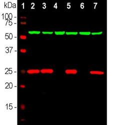
- Western blot analysis of PGP9.5 in rat brain, mouse brain, NIH-3T3, HEK293, HeLa, SH-SY5Y cells. Samples were incubated in PGP9.5 polyclonal antibody (Product # PA1-46205 using a dilution of 1:2,000. Target antibody in red, and mouse mAb to HSP60, dilution 1:10,000, in green: [1] protein standard, [2] rat brain, [3] mouse brain, [4] NIH-3T3, [5] HEK293, [6] HeLa, [7] SH-SY5Y cells. The single band at 24 kDa corresponds to the UCHL1 protein, while the 60 kDa band represents HSP60 protein. UCHL1 is detectable in CNS extracts and cells with neuronal properties but not in HeLa, NIH-3T3 and other non-neuronal cells.

- Submitted by
- Invitrogen Antibodies (provider)
- Main image
- Experimental details
- Western blot analysis of PGP9.5 in whole bovine brain extract. Samples were incubated in PGP9.5 polyclonal antibody (Product # PA1-46205). Strong and clean band at about 24 kDa.

- Submitted by
- Invitrogen Antibodies (provider)
- Main image

- Experimental details
- Western blot analysis of PGP9.5 in 0.05 mg/mL Human Brain lysate. Samples were incubated in PGP9.5 polyclonal antibody (Product # PA1-46205). This experiment was performed under reducing conditions using the 12-230 kDa separation system.

- Submitted by
- Invitrogen Antibodies (provider)
- Main image
- Experimental details
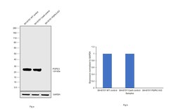
- Western blot analysis of PGP9.5/UCHL1 was performed by loading 20 µg of SH-SY5Y wild type (Lane 1), SH-SY5Y Cas9 control (Lane 2), SH-SY5Y PGP9.5/UCHL1 knockout (Lane 3) whole cell extracts. The blot was probed with Anti-PGP9.5/ UCHL1 Polyclonal Antibody (Product # PA1-46205) (1:2000 dilution) and Goat anti-Rabbit IgG (H+L), Superclonal™ Recombinant Secondary Antibody, HRP (Product # A27036) (1:4000 dilution). Loss of signal upon CRISPR mediated knockout (KO) confirms that antibody is specific to PGP9.5/UCHL1.

Supportive validation
- Submitted by
- Invitrogen Antibodies (provider)
- Main image
- Experimental details
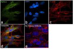
- Immunofluorescence analysis of PGP9.5 was performed using 70% confluent log phase SH-SY5Y cells. The cells were fixed with 4% paraformaldehyde for 10 minutes, permeabilized with 0.1% Triton™ X-100 for 10 minutes, and blocked with 1% BSA for 1 hour at room temperature. The cells were labeled with PGP9.5 Rabbit Polyclonal Antibody (Product # PA1-46205) at 1:500 dilution in 0.1% BSA and incubated overnight at 4 degree and then labeled with Goat anti-Mouse IgG (H+L) Superclonal™ Secondary Antibody, Alexa Fluor® 488 conjugate (Product # A27034) at a dilution of 1:2000 for 45 minutes at room temperature (Panel a: green). Nuclei (Panel b: blue) were stained with SlowFade® Gold Antifade Mountant with DAPI (Product # S36938). F-actin (Panel c: red) was stained with Rhodamine Phalloidin (Product # R415, 1:300). Panel d represents the merged image showing and cytoplasmic localization. Panel e represents control cells with no primary antibody to assess background. The images were captured at 60X magnification.

- Submitted by
- Invitrogen Antibodies (provider)
- Main image

- Experimental details
- Immunocytochemistry analysis of PGP9.5 in SH-SY5Y cells. Samples were incubated in PGP9.5 polyclonal antibody (Product # PA1-46205) using a dilution of 1:1000. This antibody in green, and costained with mouse mAb to fibrillarin, dilution 1:1,000 in red. Blue is DAPI staining of nuclear DNA. The UCHL1 antibody produces strong staining of the cellular cytoplasm of these cells which share many properties with neurons, while the fibrilarin antibody specifically labels nucleoli.

- Submitted by
- Invitrogen Antibodies (provider)
- Main image

- Experimental details
- Immunocytochemistry analysis of PGP9.5 in mixed neuron/glial cultures. Samples were incubated in PGP9.5 polyclonal antibody (Product # PA1-46205). UCHL1 antibody (green). Blue is a DNA stain. Note that the UCHL1 stains neurons strongly and specifically, and that the staining is concentrated in the cell bodies, though some does extend into the dendrites also.

- Submitted by
- Invitrogen Antibodies (provider)
- Main image

- Experimental details
- Immunofluorescence analysis of PGP9.5 was performed using 70% confluent log phase SH-SY5Y cells. The cells were fixed with 4% paraformaldehyde for 10 minutes, permeabilized with 0.1% Triton™ X-100 for 10 minutes, and blocked with 1% BSA for 1 hour at room temperature. The cells were labeled with PGP9.5 Rabbit Polyclonal Antibody (Product # PA1-46205) at 1:500 dilution in 0.1% BSA and incubated overnight at 4 degree and then labeled with Goat anti-Mouse IgG (H+L) Superclonal™ Secondary Antibody, Alexa Fluor® 488 conjugate (Product # A27034) at a dilution of 1:2000 for 45 minutes at room temperature (Panel a: green). Nuclei (Panel b: blue) were stained with SlowFade® Gold Antifade Mountant with DAPI (Product # S36938). F-actin (Panel c: red) was stained with Rhodamine Phalloidin (Product # R415, 1:300). Panel d represents the merged image showing and cytoplasmic localization. Panel e represents control cells with no primary antibody to assess background. The images were captured at 60X magnification.
