Explore
 Validate
 Learn
Learn Western blot
Antibody data
- Antibody Data
- Antigen structure
- References [1]
- Comments [0]
- Validations
- Western blot [4]
- Immunocytochemistry [1]
- Immunohistochemistry [1]
- Flow cytometry [1]
- Other assay [1]
Submit
Validation data
Reference
Comment
Report error
- Product number
- PA5-16825 - Provider product page

- Provider
- Invitrogen Antibodies
- Product name
- PGP9.5 Polyclonal Antibody
- Antibody type
- Polyclonal
- Antigen
- Recombinant full-length protein
- Description
- PA5-16825 targets Protein Gene Product 9.5 in IHC (P) applications and shows reactivity with Human samples.
- Concentration
- 0.24 mg/mL
Submitted references Schwann Cell-Derived CCL2 Promotes the Perineural Invasion of Cervical Cancer.
Huang T, Fan Q, Wang Y, Cui Y, Wang Z, Yang L, Sun X, Wang Y
Frontiers in oncology 2020;10:19
Frontiers in oncology 2020;10:19
No comments: Submit comment
Supportive validation
- Submitted by
- Invitrogen Antibodies (provider)
- Main image
- Experimental details
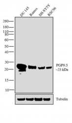
- Western blot analysis was performed on membrane enriched extracts (30 µglysate) of DU 145 (Lane 1), Ramos (Lane 2), SH-SY5Y (Lane 3) and RSC96 (Lane 4).The blots were probed with PGP9.5 Rabbit polyclonal Antibody (Product # PA5-16825, 2 µg/mL) and detected by chemiluminescence using Goat anti-Rabbit IgG (H+L) Superclonal™ Secondary Antibody, HRP conjugate (Product # A27036, 0.4 µg/mL, 1:2500 dilution). A 25 kDa band corresponding to PGP9.5 was observed across the cell lines tested. Known quantity of protein samples were electrophoresed using Novex® NuPAGE® 10 % Bis-Tris gel (Product # NP0301BOX), XCell SureLock™ Electrophoresis System (Product # EI0002) and Novex® Sharp Pre-Stained Protein Standard (Product # LC5800). Resolved proteins were then transferred onto a nitrocellulose membrane with iBlot® 2 Dry Blotting System (Product # IB21001). The membrane was probed with the relevant primary and secondary Antibody following blocking with 5% skimmed milk. Chemiluminescent detection was performed using Pierce™ ECL Western Blotting Substrate (Product # 32106).

- Submitted by
- Invitrogen Antibodies (provider)
- Main image
- Experimental details
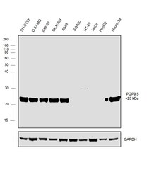
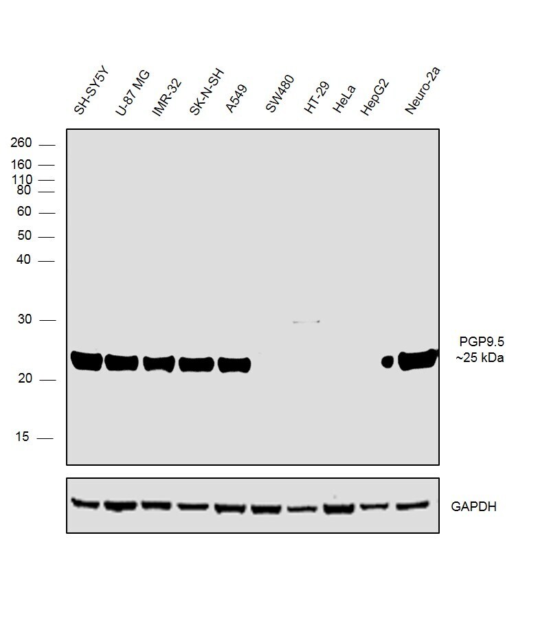
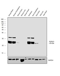
- Western blot was performed using Anti-PGP9.5 Polyclonal Antibody, (Product # PA5-16825) and a ~25 kDa band corresponding to PGP9.5 was observed across the panel tested except SW480, HT-29, HeLa and HepG2 which are reported to be negative. Whole cell extracts (30 µg lysate) of SH-SY5Y (Lane 1), U-87 MG (Lane 2), IMR-32 (Lane 3), SK-N-SH (Lane 4), A549 (Lane 5), SW480 (Lane 6), HT-29 (Lane 7), HeLa (Lane 8), HepG2 (Lane 9) and Neuro-2a (Lane 10) were electrophoresed using Novex® NuPAGE® 12% % Bis-Tris gel (Product # NP0341BOX). Resolved proteins were then transferred onto a nitrocellulose membrane (Product # IB23001) by iBlot® 2 Dry Blotting System (Product # IB21001). The blot was probed with the primary antibody (1:1000 dilution) and detected by chemiluminescence with Goat anti-Rabbit IgG (H+L) Superclonal™ Recombinant Secondary Antibody, HRP (Product # A27036, 1:4000 dilution) using the iBright FL 1000 (Product # A32752). Chemiluminescent detection was performed using Novex® ECL Chemiluminescent Substrate Reagent Kit (Product # WP20005)..
- Submitted by
- Invitrogen Antibodies (provider)
- Main image
- Experimental details
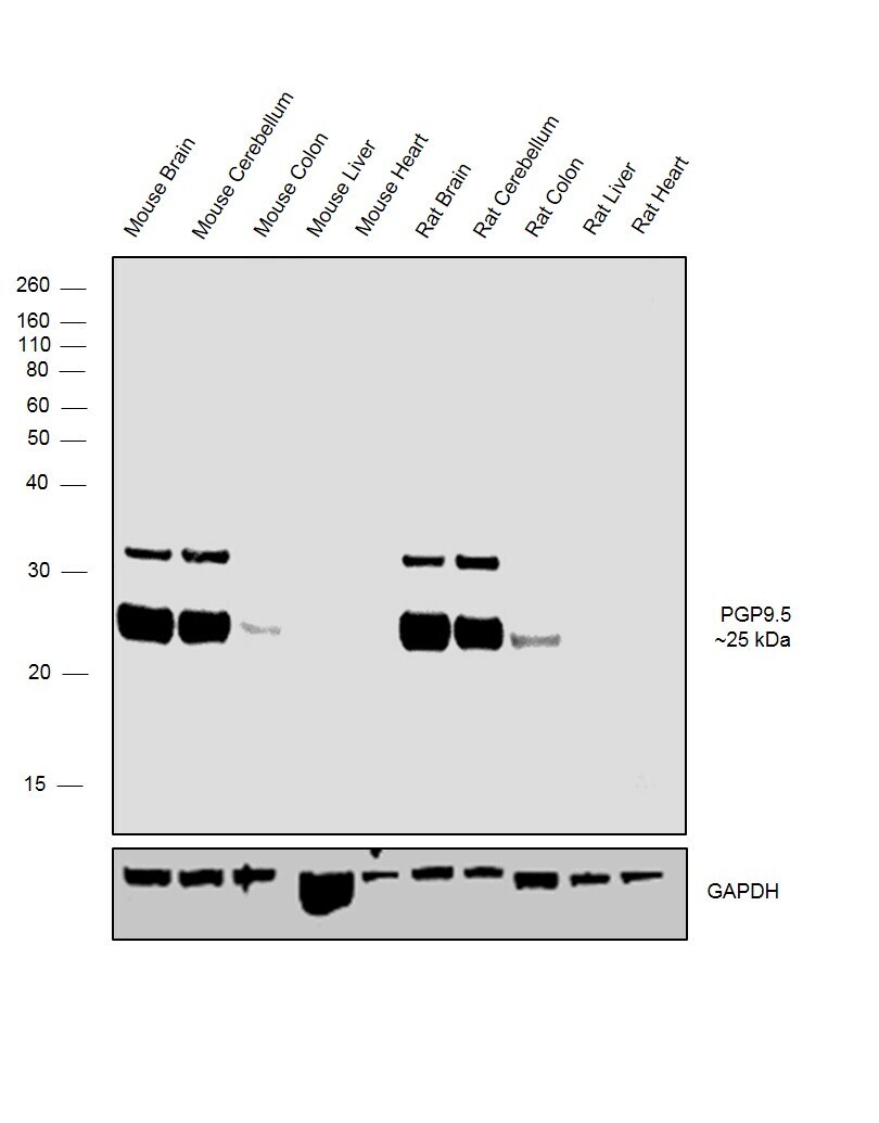
- Western blot was performed using Anti-PGP9.5 Polyclonal antibody, (Product # PA5-16825) and a ~25 kDa band corresponding to PGP9.5 was observed across the Mouse and Rat tissues except Mouse and Rat Liver and Heart which are reported to be negative. An uncharacterized band (*) at ~ 32 kDa was also observed in the PGP9.5 expressing tissues. Tissue extracts (30 µg lysate) of Mouse Brain (Lane1), Mouse Cerebellum (Lane 2), Mouse Colon (Lane 3), Mouse Liver (Lane 4), Mouse Heart (Lane 5) , Rat Brain (Lane 6), Rat Cerebellum (Lane 7), Rat Colon (Lane 8), Rat Liver (Lane 9) and Rat Heart (Lane 10) were electrophoresed using Novex® NuPAGE® 12 % Bis-Tris gel (Product # NP0341BOX). Resolved proteins were then transferred onto a nitrocellulose membrane (Product # IB23001) by iBlot® 2 Dry Blotting System (Product # IB21001). The blot was probed with the primary antibody (1:1000 dilution) and detected by chemiluminescence with Goat anti-Rabbit IgG (H+L) Superclonal™ Recombinant Secondary Antibody, HRP (Product # A27036, 1:4000 dilution) using the iBright FL 1000 (Product # A32752). Chemiluminescent detection was performed using Novex® ECL Chemiluminescent Substrate Reagent Kit (Product # WP20005)..
- Submitted by
- Invitrogen Antibodies (provider)
- Main image
- Experimental details
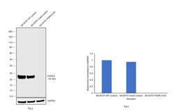
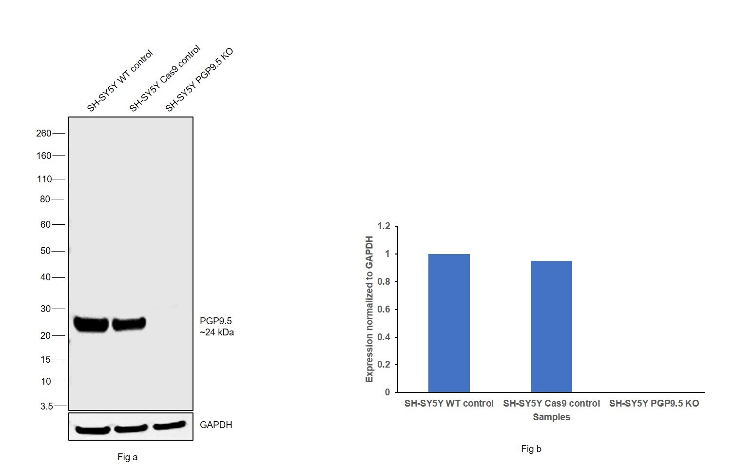
- Western blot analysis of PGP9.5/UCHL1 was performed by loading 20 µg of SH-SY5Y wild type (Lane 1), SH-SY5Y Cas9 control (Lane 2), SH-SY5Y PGP9.5/UCHL1 knockout (Lane 3) whole cell extracts. The blot was probed with Anti-PGP9.5/ UCHL1 Polyclonal Antibody (Product # PA5-16825) (1:5000 dilution) and Goat anti-Rabbit IgG (H+L), Superclonal™ Recombinant Secondary Antibody, HRP (Product # A27036) (1:4000 dilution). Loss of signal upon CRISPR mediated knockout (KO) confirms that antibody is specific to PGP9.5/UCHL1.
Supportive validation
- Submitted by
- Invitrogen Antibodies (provider)
- Main image
- Experimental details
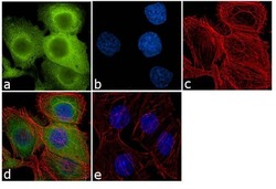
- Immunofluorescence analysis of Protein Gene Product 9.5 was performed using 70% confluent log phase DU-145 cells. The cells were fixed with 4% paraformaldehyde for 10 minutes, permeabilized with 0.1% Triton™ X-100 for 10 minutes, and blocked with 1% BSA for 1 hour at room temperature. The cells were labeled with PGP9.5 Rabbit Polyclonal Antibody (Product # PA5-16825) at 2µg/mL in 0.1% BSA and incubated for 3 hours at room temperature and then labeled with Goat anti-Rabbit IgG (H+L) Superclonal™ Secondary Antibody, Alexa Fluor® 488 conjugate (Product # A27034) at a dilution of 1:2000 for 45 minutes at room temperature (Panel a: green). Nuclei (Panel b: blue) were stained with SlowFade® Gold Antifade Mountant with DAPI (Product # S36938). F-actin (Panel c: red) was stained with Alexa Fluor® 555 Rhodamine Phalloidin (Product # R415, 1:300). Panel d represents the merged image showing cytoplasmic localization. Panel e shows the no primary antibody control. The images were captured at 60X magnification.
Supportive validation
- Submitted by
- Invitrogen Antibodies (provider)
- Main image

- Experimental details
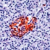
- Formalin-fixed, paraffin-embedded human pancreas stained with PGP9.5 using peroxidase-conjugate and AEC chromogen. Note staining of the islets of Langerhans.
Supportive validation
- Submitted by
- Invitrogen Antibodies (provider)
- Main image
- Experimental details
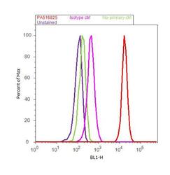
- Flow cytometry analysis of PGP9.5 was done on DU 145 cells. Cells were fixed with 70% ethanol for 10 minutes, permeabilized with 0.25% Triton™ X-100 for 20 minutes, and blocked with 5% BSA for 30 minutes at room temperature. Cells were labeled with PGP9.5 Rabbit Polyclonal Antibody (PA516825, red histogram) or with rabbit isotype control (pink histogram) at 3-5 ug/million cells in 2.5% BSA. After incubation at room temperature for 2 hours, the cells were labeled with Alexa Fluor® 488 Goat Anti-Rabbit Secondary Antibody (A11008) at a dilution of 1:400 for 30 minutes at room temperature. The representative 10,000 cells were acquired and analyzed for each sample using an Attune® Acoustic Focusing Cytometer. The purple histogram represents unstained control cells and the green histogram represents no-primary-antibody control..

Supportive validation
- Submitted by
- Invitrogen Antibodies (provider)
- Main image

- Experimental details
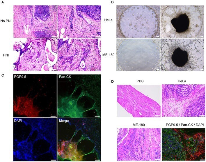
- Figure 1 The presence of perineural invasion in vitro and in vivo . (A) Hematoxylin and eosin (HE) stained image of non-PNI and PNI cervical cancer tissues. The area of nerves is marked by a dashed line. N refers to nerves and C refers to cancer cells. (B) Cervical cancer cell lines, HeLa and ME-180, induced PNI in an in vitro model. DRG was placed in the center of Matrigel (50x magnification, scale bar, 100 mum). (C) Double immunofluorescence staining of neurites and HeLa cells in the perineural niche. Staining: PGP9.5, pan-cytokeratin (pan-CK), DAPI, and overlay respectively (100x magnification, scale bar, 100 mum). (D) H&E stained image of sciatic nerves injected with PBS, HeLa and ME-180, respectively. Image in the right refers to double immunofluorescence staining of sciatic nerve (PGP9.5) and HeLa cells (pan-CK) (200x magnification, scale bar, 50 mum).