Explore
 Validate
 Learn
Learn Western blot
Antibody data
- Antibody Data
- Antigen structure
- References [3]
- Comments [0]
- Validations
- Western blot [8]
- Immunocytochemistry [1]
- Immunohistochemistry [5]
- Other assay [1]
Submit
Validation data
Reference
Comment
Report error
- Product number
- PA5-29012 - Provider product page

- Provider
- Invitrogen Antibodies
- Product name
- PGP9.5 Polyclonal Antibody
- Antibody type
- Polyclonal
- Antigen
- Synthetic peptide
- Description
- Recommended positive controls: 293T, A549, H1299, Neuro2A, GL261, rat brain, PC-12, mouse brain.
- Concentration
- 0.26 mg/mL
Submitted references SARM1 Suppresses Axon Branching Through Attenuation of Axonal Cytoskeletal Dynamics.
Evidence of Altered Peripheral Nerve Function in a Rodent Model of Diet-Induced Prediabetes.
A disease mutation reveals a role for NaV1.9 in acute itch.
Ketschek A, Holland SM, Gallo G
Frontiers in molecular neuroscience 2022;15:726962
Frontiers in molecular neuroscience 2022;15:726962
Evidence of Altered Peripheral Nerve Function in a Rodent Model of Diet-Induced Prediabetes.
Hossain MJ, Kendig MD, Wild BM, Issar T, Krishnan AV, Morris MJ, Arnold R
Biomedicines 2020 Aug 28;8(9)
Biomedicines 2020 Aug 28;8(9)
A disease mutation reveals a role for NaV1.9 in acute itch.
Salvatierra J, Diaz-Bustamante M, Meixiong J, Tierney E, Dong X, Bosmans F
The Journal of clinical investigation 2018 Dec 3;128(12):5434-5447
The Journal of clinical investigation 2018 Dec 3;128(12):5434-5447
No comments: Submit comment
Supportive validation
- Submitted by
- Invitrogen Antibodies (provider)
- Main image

- Experimental details
- Western blot analysis of PGP9.5 using 30 µg of A) A549 and B) H1299 lysate. Samples were loaded onto a 12% SDS-PAGE gel and probed with a PGP9.5 polyclonal antibody (Product # PA5-29012) at a dilution of 1:5000.
- Submitted by
- Invitrogen Antibodies (provider)
- Main image

- Experimental details
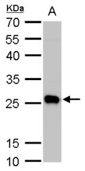
- Western blot analysis of PGP9.5 using 50 µg mouse brain lysate. Samples were loaded onto a 12% SDS-PAGE gel and probed with a PGP9.5 polyclonal antibody (Product # PA5-29012) at a dilution of 1:5000.
- Submitted by
- Invitrogen Antibodies (provider)
- Main image

- Experimental details
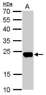
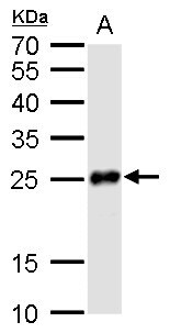
- Western blot analysis of PGP9.5 using 50 µg rat brain lysate. Samples were loaded onto a 12% SDS-PAGE gel and probed with a PGP9.5 polyclonal antibody (Product # PA5-29012) at a dilution of 1:10,000.

- Submitted by
- Invitrogen Antibodies (provider)
- Main image

- Experimental details
- Western Blot analysis of PGP9.5 was performed by separating 50 µg of rat tissue extract by 12% SDS-PAGE. Proteins were transferred to a membrane and probed with a PGP9.5 Polyclonal Antibody (Product # PA5-29012) at a dilution of 1:10,000.

- Submitted by
- Invitrogen Antibodies (provider)
- Main image
- Experimental details
- Western Blot analysis of PGP9.5 was performed by separating 30 µg of whole cell lysates by 12% SDS-PAGE. Proteins were transferred to a membrane and probed with a PGP9.5 Polyclonal Antibody (Product # PA5-29012) at a dilution of 1:10000. The HRP-conjugated anti-rabbit IgG antibody was used to detect the primary antibody. A. Neuro2A, B. GL261.

- Submitted by
- Invitrogen Antibodies (provider)
- Main image
- Experimental details
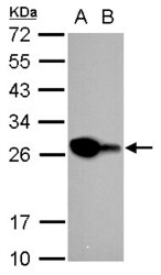
- Western Blot analysis of PGP9.5 was performed by separating 50 µg of mouse brain extracts by 12 % SDS-PAGE. Proteins were transferred to a membrane and probed with a PGP9.5 Polyclonal Antibody (Product # PA5-29012) at a dilution of 1:5000.

- Submitted by
- Invitrogen Antibodies (provider)
- Main image

- Experimental details
- Western Blot analysis of PGP9.5 was performed by separating 50 µg of rat brain extracts by 12 % SDS-PAGE. Proteins were transferred to a membrane and probed with a PGP9.5 Polyclonal Antibody (Product # PA5-29012) at a dilution of 1:10000.
- Submitted by
- Invitrogen Antibodies (provider)
- Main image

- Experimental details
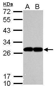
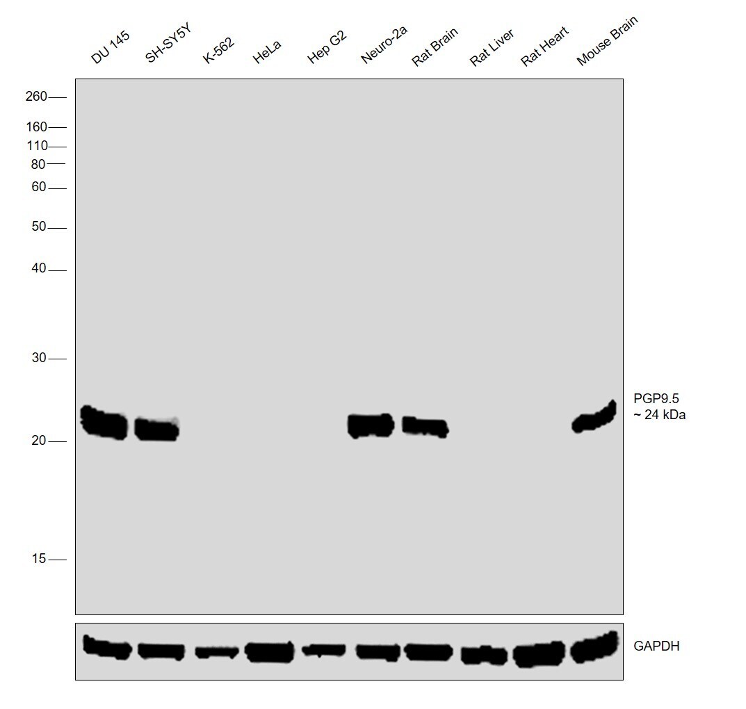
- Western blot was performed using Anti-PGP9.5 Polyclonal Antibody (Product # PA5-29012) and a 24 kDa band corresponding to PGP9.5 was observed across DU 145, SH-SY5Y, Neuro-2a, Rat Brain and Mouse Brain. Whole Cell Extract-WCL (30 µg lysate) of DU 145 (Lane 1), SH-SY5Y (Lane 2), K-562 (Lane 3), HeLa (Lane 4), Hep G2 (Lane 5), Neuro-2a (Lane 6), Rat Brain (Lane 7), Rat Liver (Lane 8), Rat Heart (Lane 9) and Mouse Brain (Lane 10) were electrophoresed using NuPAGE™ 12% Bis-Tris Protein Gel (Product # NP0342BOX). Resolved proteins were then transferred onto a Nitrocellulose membrane (Product # IB23002) by iBlot® 2 Dry Blotting System (Product # IB21001). The blot was probed with the primary antibody (1:2500 dilution) and detected by chemiluminescence with Goat anti-Rabbit IgG (H+L) Superclonal™ Recombinant Secondary Antibody, HRP (Product # A27036, 1:4000 dilution) using the iBright FL 1000 (Product # A32752). Chemiluminescent detection was performed using Novex® ECL Chemiluminescent Substrate Reagent Kit (Product # WP20005).
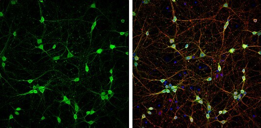
Supportive validation
- Submitted by
- Invitrogen Antibodies (provider)
- Main image
- Experimental details
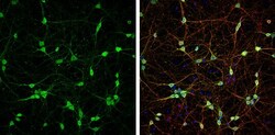
- Immunocytochemistry-Immunofluorescence analysis of PGP9.5 was performed in DIV9 rat E18 primary cortical neurons fixed in 4% paraformaldehyde at RT for 15 min. Green: PGP9.5 Polyclonal Antibody (Product # PA5-29012) diluted at 1:500. Red: beta Tubulin 3/ Tuj1, stained by beta Tubulin 3/ Tuj1 antibody. Blue: Fluoroshield with DAPI.
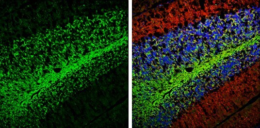
Supportive validation
- Submitted by
- Invitrogen Antibodies (provider)
- Main image
- Experimental details
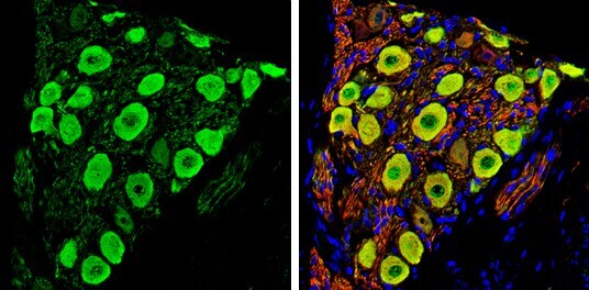
- Immunohistochemistry (Frozen) analysis of PGP9.5 was performed in frozen sectioned E13.5 Rat brain tissue using PGP9.5 Polyclonal Antibody (Product # PA5-29012) at a dilution of 1:250 (Green). Red: beta Tubulin 3/ TUJ1, a mature neuron marker, stained by beta Tubulin 3/ TUJ1 antibody diluted at 1:500. Blue: Fluoroshield with DAPI.

- Submitted by
- Invitrogen Antibodies (provider)
- Main image

- Experimental details
- Immunohistochemistry (Frozen) analysis of PGP9.5 was performed in frozen-sectioned adult mouse cerebellum tissue using PGP9.5 Polyclonal Antibody (Product # PA5-29012) at a dilution of 1:250 (Green). Red: beta Tubulin 3/ TUJ1, stained by beta Tubulin 3/ TUJ1 antibody diluted at 1:500. Blue: Fluoroshield with DAPI.
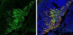
- Submitted by
- Invitrogen Antibodies (provider)
- Main image
- Experimental details
- Immunohistochemistry (Paraffin) analysis of PGP9.5 was performed in paraffin-embedded mouse skin tissue. Green: PGP9.5 stained by PGP9.5 Polyclonal Antibody (Product # PA5-29012) at a dilution of 1:250. Red: beta Tubulin 3/ Tuj1, a marker, stained by beta Tubulin 3/ Tuj1 antibody. Blue: Fluoroshield with DAPI. Antigen Retrieval: Citrate buffer, pH 6.0, 15 min.

- Submitted by
- Invitrogen Antibodies (provider)
- Main image

- Experimental details
- Immunohistochemistry (Paraffin) analysis of PGP9.5 was performed in paraffin-embedded rat colon tissue. Green: PGP9.5 stained by PGP9.5 Polyclonal Antibody (Product # PA5-29012) at a dilution of 1:250. Red: beta Tubulin 3/ Tuj1, a marker, stained by beta Tubulin 3/ Tuj1 antibody. Blue: Fluoroshield with DAPI. Antigen Retrieval: Citrate buffer, pH 6.0, 15 min.
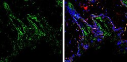
- Submitted by
- Invitrogen Antibodies (provider)
- Main image
- Experimental details
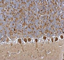
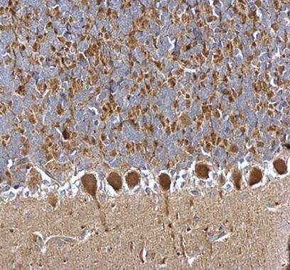
- PGP9.5 Polyclonal Antibody detects PGP9.5 protein at cytosol on rat hind brain by immunohistochemical analysis. Sample: Paraffin-embedded rat hind brain. PGP9.5 Polyclonal Antibody (Product # PA5-29012) dilution: 1:500. Antigen Retrieval: EDTA based buffer, pH 8.0, 15 min.
Supportive validation
- Submitted by
- Invitrogen Antibodies (provider)
- Main image
- Experimental details
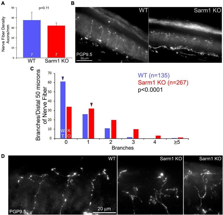
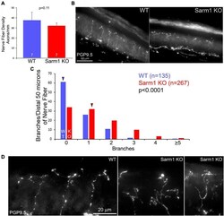
- FIGURE 3 Terminals of SARM1 KO sensory cutaneous axons exhibit more complex types of morphology than those of WT axons in vivo . (A) Graph of the intra-epidermal nerve fiber density in WT and SARM1 KO skin. n = number of mice (120-127 days of age). (B) Examples of WT and SARM1 KO skin sections stained with anti-PGP9.5 used to obtain the intra-epidermal nerve fiber density measurements presented in (A) . (C) Histogram of the number of branches present along the distal 50 microns of sensory fibers. A value of 0 reflects a fiber with no branches and only its terminus. Arrowheads denote the medians. (D) Examples of the morphology of the termini of nerve fibers in WT and SARM1 KO samples.