 Explore
 Validate
 Learn
Learn Western blot
 Immunocytochemistry
Antibody data
- Antibody Data
- Antigen structure
- References [3]
- Comments [0]
- Validations
- Western blot [1]
Submit
Validation data
Reference
Comment
Report error
- Product number
- NB110-58872 - Provider product page

- Provider
- Novus Biologicals
- Proper citation
- Novus Cat#NB110-58872, RRID:AB_877619
- Product name
- Chicken Polyclonal UCH-L1/PGP9.5 Antibody
- Antibody type
- Polyclonal
- Description
- Ammonium sulfate precipitation.
- Reactivity
- Human, Mouse, Rat, Bovine, Porcine
- Host
- Chicken/Avian
- Isotype
- IgY
- Vial size
- 0.1 ml
- Storage
- Store at 4C short term. Aliquot and store at -20C long term. Avoid freeze-thaw cycles.
Submitted references Synergistic regulation of serotonin and opioid signaling contributes to pain insensitivity in Nav1.7 knockout mice.
Subgroup-elimination transcriptomics identifies signaling proteins that define subclasses of TRPV1-positive neurons and a novel paracrine circuit.
Pain modulators regulate the dynamics of PKA-RII phosphorylation in subgroups of sensory neurons.
Isensee J, Krahé L, Moeller K, Pereira V, Sexton JE, Sun X, Emery E, Wood JN, Hucho T
Science signaling 2017 Jan 10;10(461)
Science signaling 2017 Jan 10;10(461)
Subgroup-elimination transcriptomics identifies signaling proteins that define subclasses of TRPV1-positive neurons and a novel paracrine circuit.
Isensee J, Wenzel C, Buschow R, Weissmann R, Kuss AW, Hucho T
PloS one 2014;9(12):e115731
PloS one 2014;9(12):e115731
Pain modulators regulate the dynamics of PKA-RII phosphorylation in subgroups of sensory neurons.
Isensee J, Diskar M, Waldherr S, Buschow R, Hasenauer J, Prinz A, Allgöwer F, Herberg FW, Hucho T
Journal of cell science 2014 Jan 1;127(Pt 1):216-29
Journal of cell science 2014 Jan 1;127(Pt 1):216-29
No comments: Submit comment
Supportive validation
- Submitted by
- Novus Biologicals (provider)
- Main image
- Experimental details
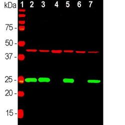
- Western Blot: UCH-L1/PGP9.5 Antibody [NB110-58872] - Analysis of equal amounts of different tissue and cell lysates using chicken pAb to UCHL1, NB110-58872, dilution 1:2,000 in green, and mouse mAb to Actin, dilution 1:1,000, in red: [1] protein standard, [2] rat brain, [3] mouse brain, [4] NIH-3T3, [5] HEK293, [6] HeLa and [7] SH-SY5Y cells. The single band at 24 kDa mark corresponds to UCHL1 protein which is detectable in CNS extracts and lysates of cells with neuronal properties but not in lysates of HeLa, NIH-3T3 and other non-neuronal cells. Actin is detected with apparent molecular weight of 42 kDa and provides an excellent loading control.
