 Explore
 Validate
 Learn
Learn Western blot
Antibody data
- Antibody Data
- Antigen structure
- References [2]
- Comments [0]
- Validations
- Western blot [4]
- Immunocytochemistry [1]
- Immunohistochemistry [1]
- Other assay [2]
Submit
Validation data
Reference
Comment
Report error
- Product number
- PA5-34901 - Provider product page

- Provider
- Invitrogen Antibodies
- Product name
- ALDH1A1 Polyclonal Antibody
- Antibody type
- Polyclonal
- Antigen
- Recombinant protein fragment
- Description
- Recommended positive controls: A549, mouse liver, rat liver, Huh7, HepG2.
- Concentration
- 1 mg/mL
Submitted references Parathyroid Hormone-Related Protein Inhibition Blocks Triple-Negative Breast Cancer Expansion in Bone Through Epithelial to Mesenchymal Transition Reversal.
Low P16(INK4A) Expression Associated with High Expression of Cancer Stem Cell Markers Predicts Poor Prognosis in Cervical Cancer after Radiotherapy.
Li J, Camirand A, Zakikhani M, Sellin K, Guo Y, Luan X, Mihalcioiu C, Kremer R
JBMR plus 2022 Jun;6(6):e10587
JBMR plus 2022 Jun;6(6):e10587
Low P16(INK4A) Expression Associated with High Expression of Cancer Stem Cell Markers Predicts Poor Prognosis in Cervical Cancer after Radiotherapy.
Fu HC, Chuang IC, Yang YC, Chuang PC, Lin H, Ou YC, Chang Chien CC, Huang HS, Kang HY
International journal of molecular sciences 2018 Aug 27;19(9)
International journal of molecular sciences 2018 Aug 27;19(9)
No comments: Submit comment
Supportive validation
- Submitted by
- Invitrogen Antibodies (provider)
- Main image

- Experimental details
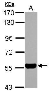
- Western Blot using ALDH1A1 Polyclonal Antibody (Product # PA5-34901). Sample (30 µg of whole cell lysate). Lane A: A549. 7.5% SDS PAGE. ALDH1A1 Polyclonal Antibody (Product # PA5-34901) diluted at 1:10,000. The HRP-conjugated anti-rabbit IgG antibody was used to detect the primary antibody.
- Submitted by
- Invitrogen Antibodies (provider)
- Main image

- Experimental details
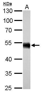
- ALDH1A1 Polyclonal Antibody detects ALDH1A1 protein by western blot analysis. A. 50 µg mouse liver lysate/extract.10% SDS-PAGE. ALDH1A1 Polyclonal Antibody (Product # PA5-34901) dilution: 1:5,000. The HRP-conjugated anti-rabbit IgG antibody was used to detect the primary antibody.
- Submitted by
- Invitrogen Antibodies (provider)
- Main image
- Experimental details
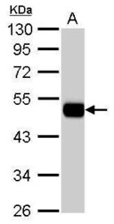
- ALDH1A1 Polyclonal Antibody detects ALDH1A1 protein by western blot analysis. A. 50 µg rat liver lysate/extract.10% SDS-PAGE. ALDH1A1 Polyclonal Antibody (Product # PA5-34901) dilution: 1:5,000. The HRP-conjugated anti-rabbit IgG antibody was used to detect the primary antibody.

- Submitted by
- Invitrogen Antibodies (provider)
- Main image

- Experimental details
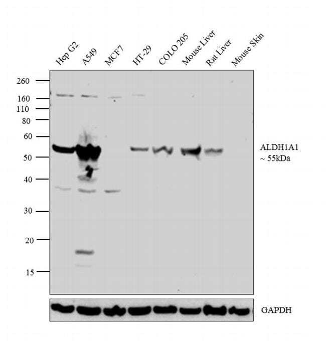
- Western blot analysis was performed on whole cell extracts (30 µg lysate) of Hep G2 (Lane 1), A549 (Lane 2), MCF7 (Lane 3), HT-29 (Lane 4), COLO 205 (Lane 5), tissue extract of Mouse Liver (Lane 6), Rat Liver (Lane 7) and Mouse Skin (Lane 8). The blot was probed with Anti-ALDH1A1 Polyclonal Antibody (Product # PA5-34901, 1:1000 dilution) and detected by chemiluminescence using Goat anti-Rabbit IgG (H+L) Superclonal™ Secondary Antibody, HRP conjugate (Product # A27036, 0.25 µg/ml, 1:4000 dilution). A 55 kDa band corresponding to ALDH1A1 was detected across the cell lines and tissues tested except for MCF7 and Mouse Skin which are reported negative for ALDH1A1 expression.
Supportive validation
- Submitted by
- Invitrogen Antibodies (provider)
- Main image

- Experimental details
- Immunofluorescent analysis of ALDH1A1 in methanol-fixed A549 cells using an ALDH1A1 polyclonal antibody (Product # PA5-34901) at a 1:500 dilution.

Supportive validation
- Submitted by
- Invitrogen Antibodies (provider)
- Main image

- Experimental details
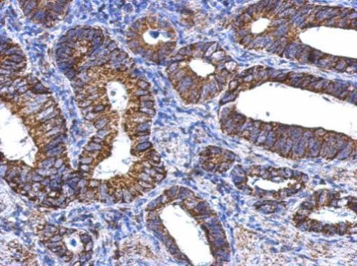
- Immunohistochemical analysis of paraffin-embedded human gastric cancer, using ALDH1A1 (Product # PA5-34901) antibody at 1:500 dilution. Antigen Retrieval: EDTA based buffer, pH 8.0, 15 min.
Supportive validation
- Submitted by
- Invitrogen Antibodies (provider)
- Main image

- Experimental details
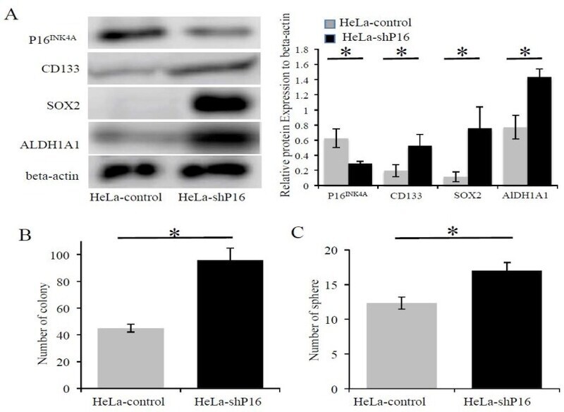
- Figure 5 The inhibition of P16 INK4A protein expression increased SOX2, ALDH1A1 expression, and self-renewal ability in cervical cancer cells. ( A ) Expression of stem cell markers (CD133, SOX2, and ALDH1A1) at the protein level in the HeLa-control and HeLa-shP16 cells were detected by Western blot and relative protein expression to beta-actin protein was shown. ( B ) Colony formation assay of HeLa-control and HeLa-shP16 cells were used to evaluate the ability of self-renew. ( C ) The sphere formation assay was used to measure the anchorage-independent growth of HeLa-control and HeLa-shP16 cells in ultra-low attachment plates. Each bar represents the mean +- SD of three independent experiments. Student's t -test was used for continuous variables between the two groups. * p < 0.05. Data are presented as the mean +- SD of three independent experiments.
- Submitted by
- Invitrogen Antibodies (provider)
- Main image
- Experimental details
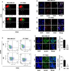
- 3 Fig Pthlh ablation reduces the CSC subpopulation in MBA-MB-231 and PT-TNBCs cells. ( A ) Flow analysis of CD44 and CD24-expressing MDA-MB-231 (left) and PT-TNBC cells (right). ( B , C ) IF staining for CD44 and CD24 in WT and KO MDA-MB-231 and PT-TNBCs. ( D ) Flow analysis of CD49f and CD24 in MDA-MB-231 (left) and PT-TNBC cells (right). ( E , F ) IF staining for ALDH1 and mRNA levels in WT (empty vector) and KO MDA-MB-231 and PT-TNBCs. DAPI: blue; CD44: red; CD24: green; ALDH1: green. n = 5, p < 0.001. Scale bars = 100 mum ( B , C ), 50 mum ( E , F ).
