 Explore
 Validate
 Learn
Learn Western blot
Antibody data
- Antibody Data
- Antigen structure
- References [0]
- Comments [0]
- Validations
- Western blot [3]
- Immunocytochemistry [1]
Submit
Validation data
Reference
Comment
Report error
- Product number
- 702728 - Provider product page

- Provider
- Invitrogen Antibodies
- Product name
- ALDH1A1 Recombinant Rabbit Monoclonal Antibody (20H2L4)
- Antibody type
- Monoclonal
- Antigen
- Synthetic peptide
- Reactivity
- Human
- Host
- Rabbit
- Isotype
- IgG
- Antibody clone number
- 20H2L4
- Vial size
- 100 µg
- Concentration
- 0.5 mg/mL
- Storage
- Store at 4°C short term. For long term storage, store at -20°C, avoiding freeze/thaw cycles.
No comments: Submit comment
Supportive validation
- Submitted by
- Invitrogen Antibodies (provider)
- Main image
- Experimental details
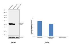
- Knockdown of ALDH1A1 was achieved by transfecting A549 cells with ALDH1A1 specific validated siRNA (Silencer® select Product # s225). Western blot analysis (Fig a) was performed using whole cell extracts from ALDH1A1 knockdown cells (Lane 3), non-specific scrambled siRNA transfected cells (Lane 2) and untransfected cells (Lane 1). The blots were probed with Anti-ALDH1A1 Recombinant Rabbit Polyclonal Antibody (Product # 702728, 1:200 dilution) and Goat anti-Rabbit IgG (H+L) Superclonal™ Secondary Antibody, HRP conjugate (Product # A27036, 0.25 µg/mL, 1:4000 dilution). Densitometric analysis of this Western blot is shown in histogram (Fig b). Loss of signal upon siRNA mediated knock down confirms that antibody is specific to ALDH1A1.

- Submitted by
- Invitrogen Antibodies (provider)
- Main image
- Experimental details
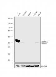
- Western blot analysis was performed on Whole cell extracts (30 µg lysate) of A549 (Lane 1), Colo 205 (Lane 2), LNCaP (Lane 3), MKN45 (Lane 4) and HT-29 (Lane 5). The blots were probed with Anti-ALDH1A1 Recombinant Rabbit Monoclonal Antibody (Product # 702728, 1:200 dilution) and detected by chemiluminescence using Goat anti-Rabbit IgG (H+L) Superclonal™ Secondary Antibody, HRP conjugate (Product # A27036, 0.25 µg/mL, 1:4000 dilution). A ~52 kDa band corresponding to ALDH1A1 was observed to be prominently present in A549 and detected in few other cell lines tested.

- Submitted by
- Invitrogen Antibodies (provider)
- Main image
- Experimental details
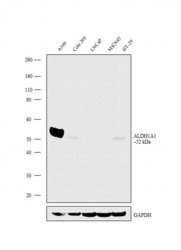
- Western blot analysis was performed on Whole cell extracts (30 µg lysate) of A549 (Lane 1), Colo 205 (Lane 2), LNCaP (Lane 3), MKN45 (Lane 4) and HT-29 (Lane 5). The blots were probed with Anti-ALDH1A1 Recombinant Rabbit Monoclonal Antibody (Product # 702728, 1:200 dilution) and detected by chemiluminescence using Goat anti-Rabbit IgG (H+L) Superclonal™ Secondary Antibody, HRP conjugate (Product # A27036, 0.25 µg/mL, 1:4000 dilution). A ~52 kDa band corresponding to ALDH1A1 was observed to be prominently present in A549 and detected in few other cell lines tested.

Supportive validation
- Submitted by
- Invitrogen Antibodies (provider)
- Main image

- Experimental details
- For immunofluorescence analysis, A549 cells were fixed and permeabilized for detection of endogenous ALDH1A1 using Anti- ALDH1A1 Recombinant Rabbit Monoclonal Antibody (Product # 702728, 1:100 dilution) and labeled with Goat anti-Rabbit IgG (H+L) Superclonal™ Secondary Antibody, Alexa Fluor® 488 conjugate (Product # A27034, 1:2000 dilution). Panel a) shows representative cells that were stained for detection and localization of ALDH1A1 protein (green), Panel b) is stained for nuclei (blue) using SlowFade® Gold Antifade Mountant with DAPI (Product # S36938). Panel c) represents cytoskeletal F-actin staining using Rhodamine Phalloidin (Product # R415, 1:300). Panel d) is a composite image of Panels a, b and c clearly demonstrating cytoplasmic localization of ALDH1A1. Panel e) represents control cells with no primary antibody to assess background. The images were captured at 60X magnification.
