Explore
 Validate
 Learn
Learn Western blot
 Immunohistochemistry
Antibody data
- Antibody Data
- Antigen structure
- References [1]
- Comments [0]
- Validations
- Immunohistochemistry [1]
Submit
Validation data
Reference
Comment
Report error
- Product number
- PA5-18483 - Provider product page

- Provider
- Invitrogen Antibodies
- Product name
- ALDH1A1 Polyclonal Antibody
- Antibody type
- Polyclonal
- Antigen
- Synthetic peptide
- Description
- This antibody is predicted to react with mouse and rat based on sequence homology. This antibody is tested in Peptide ELISA: antibody detection limit dilution 32,000.
- Reactivity
- Human
- Host
- Goat
- Isotype
- IgG
- Vial size
- 100 μg
- Concentration
- 0.5 mg/mL
- Storage
- -20°C, Avoid Freeze/Thaw Cycles
Submitted references HOXA9 Overexpression Contributes to Stem Cell Overpopulation That Drives Development and Growth of Colorectal Cancer.
Osmond B, Facey COB, Zhang C, Boman BM
International journal of molecular sciences 2022 Jun 18;23(12)
International journal of molecular sciences 2022 Jun 18;23(12)
No comments: Submit comment
Supportive validation
- Submitted by
- Invitrogen Antibodies (provider)
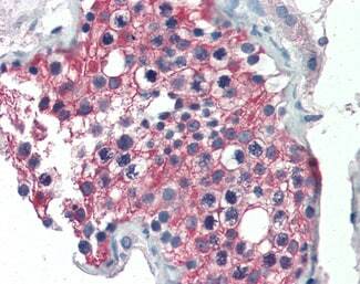
- Main image
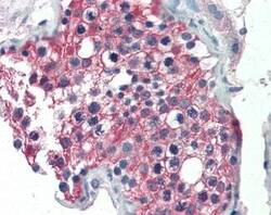
- Experimental details
- Immunohistochemistry analysis of ALDH1A1 in human testis. Samples were incubated with ALDH1A1 polyclonal antibody (Product # PA5-18483) using a dilution of 5 µg/mL. Formalin-fixed, paraffin-embedded tissue after heat-induced antigen retrieval.