Explore
 Validate
 Learn
Learn Western blot
Antibody data
- Antibody Data
- Antigen structure
- References [3]
- Comments [0]
- Validations
- Western blot [4]
- Immunocytochemistry [3]
- Immunohistochemistry [2]
- Other assay [2]
Submit
Validation data
Reference
Comment
Report error
- Product number
- PA5-32127 - Provider product page

- Provider
- Invitrogen Antibodies
- Product name
- ALDH1A1 Polyclonal Antibody
- Antibody type
- Polyclonal
- Antigen
- Recombinant full-length protein
- Description
- Recommended positive controls: A549, mouse liver, rat liver. Predicted reactivity: Mouse (90%), Rat (90%), Xenopus laevis (81%), Dog (90%), Rabbit (90%), Chicken (86%), Sheep (93%), Chimpanzee (100%), Bovine (93%). Store product as a concentrated solution. Centrifuge briefly prior to opening the vial.
- Reactivity
- Human, Mouse, Rat
- Host
- Rabbit
- Isotype
- IgG
- Vial size
- 100 μL
- Concentration
- 1 mg/mL
- Storage
- Store at 4°C short term. For long term storage, store at -20°C, avoiding freeze/thaw cycles.
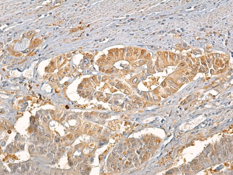
Submitted references Pathological and immunohistochemical study of colon cancer. Evaluation of markers for colon cancer stem cells.
Rhus coriaria L. (Sumac) Demonstrates Oncostatic Activity in the Therapeutic and Preventive Model of Breast Carcinoma.
Friend leukemia virus integration 1 is a predictor of poor prognosis of breast cancer and promotes metastasis and cancer stem cell properties of breast cancer cells.
Ilie DS, Mitroi G, Păun I, Ţenea-Cojan TŞ, Neamţu C, Totolici BD, Sapalidis K, Mogoantă SŞ, Murea A
Romanian journal of morphology and embryology = Revue roumaine de morphologie et embryologie 2021 Jan-Mar;62(1):117-124
Romanian journal of morphology and embryology = Revue roumaine de morphologie et embryologie 2021 Jan-Mar;62(1):117-124
Rhus coriaria L. (Sumac) Demonstrates Oncostatic Activity in the Therapeutic and Preventive Model of Breast Carcinoma.
Kubatka P, Kello M, Kajo K, Samec M, Liskova A, Jasek K, Koklesova L, Kuruc T, Adamkov M, Smejkal K, Svajdlenka E, Solar P, Pec M, Büsselberg D, Sadlonova V, Mojzis J
International journal of molecular sciences 2020 Dec 26;22(1)
International journal of molecular sciences 2020 Dec 26;22(1)
Friend leukemia virus integration 1 is a predictor of poor prognosis of breast cancer and promotes metastasis and cancer stem cell properties of breast cancer cells.
Yan X, Yu Y, Li L, Chen N, Song W, He H, Dong J, Liu X, Cui J
Cancer medicine 2018 Aug;7(8):3548-3560
Cancer medicine 2018 Aug;7(8):3548-3560
No comments: Submit comment
Supportive validation
- Submitted by
- Invitrogen Antibodies (provider)
- Main image
- Experimental details
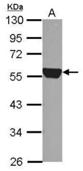
- Western Blot using ALDH1A1 Polyclonal Antibody (Product # PA5-32127). Sample (30 µg of whole cell lysate). Lane A: A549. 10% SDS PAGE. ALDH1A1 Polyclonal Antibody (Product # PA5-32127) diluted at 1:5,000. The HRP-conjugated anti-rabbit IgG antibody was used to detect the primary antibody.

- Submitted by
- Invitrogen Antibodies (provider)
- Main image

- Experimental details
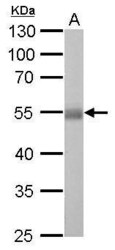
- ALDH1A1 Polyclonal Antibody detects ALDH1A1 protein by western blot analysis. A. 50 µg mouse liver lysate/extract.10% SDS-PAGE. ALDH1A1 Polyclonal Antibody (Product # PA5-32127) dilution: 1:10,000. The HRP-conjugated anti-rabbit IgG antibody was used to detect the primary antibody.

- Submitted by
- Invitrogen Antibodies (provider)
- Main image
- Experimental details
- ALDH1A1 Polyclonal Antibody detects ALDH1A1 protein by western blot analysis. A. 50 µg rat liver lysate/extract.10% SDS-PAGE. ALDH1A1 Polyclonal Antibody (Product # PA5-32127) dilution: 1:5,000. The HRP-conjugated anti-rabbit IgG antibody was used to detect the primary antibody.

- Submitted by
- Invitrogen Antibodies (provider)
- Main image

- Experimental details
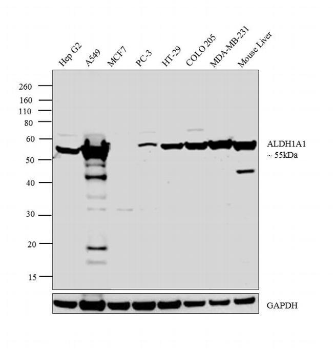
- Western blot analysis was performed on whole cell extracts (30 µg lysate) of Hep G2 (Lane 1), A549 (Lane 2), MCF7 (Lane 3), PC-3 (Lane 4), HT-29 (Lane 5), COLO 205 (Lane 6), MDA-MB-231 (Lane 7) and tissue extract of Mouse Liver (Lane 8). The blot was probed with Anti-ALDH1A1 Polyclonal Antibody (Product # PA5-32127, 1:1,000 dilution) and detected by chemiluminescence using Goat anti-Rabbit IgG (Heavy Chain) Superclonal™ Secondary Antibody, HRP conjugate (Product # A27036, 0.25 µg/mL, 1:4,000 dilution). A 55 kDa band corresponding to ALDH1A1 was detected across the cell lines and tissue tested except for MCF7 which is reported negative for ALDH1A1 expression.
Supportive validation
- Submitted by
- Invitrogen Antibodies (provider)
- Main image
- Experimental details
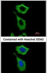
- Immunofluorescent analysis of ALDH1A1 in methanol-fixed A549 cells using an ALDH1A1 polyclonal antibody (Product # PA5-32127) at a 1:500 dilution.

- Submitted by
- Invitrogen Antibodies (provider)
- Main image
- Experimental details
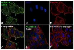
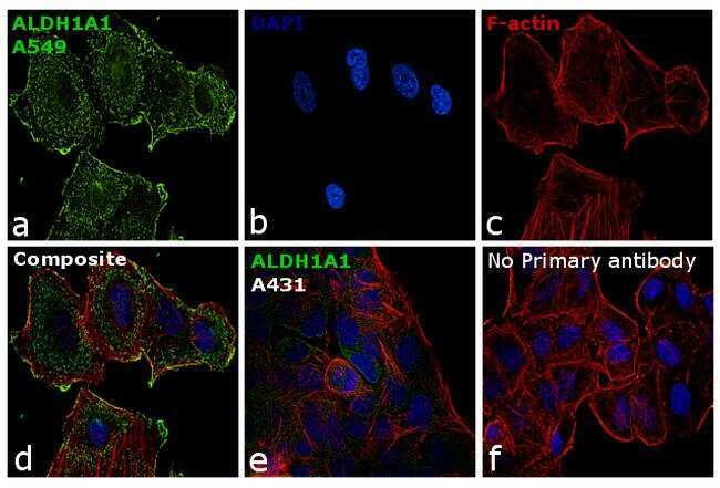
- Immunofluorescence analysis of ALDH1A1 was performed using 70% confluent log phase A549 cells. The cells were fixed with 4% paraformaldehyde for 10 minutes, permeabilized with 0.1% Triton™ X-100 for 15 minutes, and blocked with 1% BSA for 1 hour at room temperature. The cells were labeled with ALDH1A1 Polyclonal Antibody (Product # PA5-32127) at 5 µg/mL in 0.1% BSA, incubated at 4 degree Celsius overnight and then labeled with Goat anti-Rabbit IgG (H+L) Superclonal™ Secondary Antibody, Alexa Fluor® 488 conjugate (Product # A27034) at a dilution of 1:2000 for 45 minutes at room temperature (Panel a: green). Nuclei (Panel b: blue) were stained with ProLong™ Diamond Antifade Mountant with DAPI (Product # P36962). F-actin (Panel c: red) was stained with Rhodamine Phalloidin (Product # R415). Panel d represents the merged image showing cytoplasmic localization. Panel e represents A-431 cells having lower expression of ALDH1A1. Panel f represents control cells with no primary antibody to assess background. The images were captured at 60X magnification.
- Submitted by
- Invitrogen Antibodies (provider)
- Main image
- Experimental details
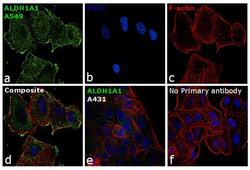
- Immunofluorescence analysis of ALDH1A1 was performed using 70% confluent log phase A549 cells. The cells were fixed with 4% paraformaldehyde for 10 minutes, permeabilized with 0.1% Triton™ X-100 for 15 minutes, and blocked with 1% BSA for 1 hour at room temperature. The cells were labeled with ALDH1A1 Polyclonal Antibody (Product # PA5-32127) at 5 µg/mL in 0.1% BSA, incubated at 4 degree Celsius overnight and then labeled with Goat anti-Rabbit IgG (Heavy Chain) Superclonal™ Secondary Antibody, Alexa Fluor® 488 conjugate (Product # A27034) at a dilution of 1:2000 for 45 minutes at room temperature (Panel a: green). Nuclei (Panel b: blue) were stained with ProLong™ Diamond Antifade Mountant with DAPI (Product # P36962). F-actin (Panel c: red) was stained with Rhodamine Phalloidin (Product # R415). Panel d represents the merged image showing cytoplasmic localization. Panel e represents A-431 cells having lower expression of ALDH1A1. Panel f represents control cells with no primary antibody to assess background. The images were captured at 60X magnification.

Supportive validation
- Submitted by
- Invitrogen Antibodies (provider)
- Main image
- Experimental details
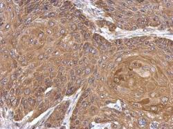
- Immunohistochemical analysis of paraffin-embedded Cal27 xenograft, using ALDH1A1 (Product # PA5-32127) antibody at 1:500 dilution. Antigen Retrieval: EDTA based buffer, pH 8.0, 15 min.

- Submitted by
- Invitrogen Antibodies (provider)
- Main image

- Experimental details
- Immunohistochemical analysis of paraffin-embedded Cal27 xenograft, using ALDH1A1 (Product # PA5-32127) antibody at 1:500 dilution. Antigen Retrieval: EDTA based buffer, pH 8.0, 15 min.

Supportive validation
- Submitted by
- Invitrogen Antibodies (provider)
- Main image
- Experimental details
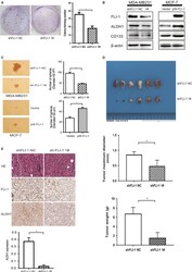
- Figure 4 Knockdown of FLI -1 inhibits clonogenicity and cancer stem cell properties in vitro and suppresses tumorigenesis in vivo. A, The colony-forming assay demonstrated that the knockdown of FLI -1 reduced the cell colony-forming number. B, The knockdown of FLI -1 decreased the expression of mammary stem cell markers ( ALDH 1 and CD 133) compared with those of the control group. The overexpression of FLI -1 in the MCF -7 cells upregulated the expression of stem cell markers. C, In the sh FLI -1 group of the MDA - MB 231 cells, the sphere-forming ability was attenuated. The overexpression of FLI -1 in the MCF -7 cells upregulated the sphere-forming capacity. Each bar represents mean +- standard deviation ( SD ). * P < .05. D, The images of excised tumors 30 days after the orthotopic injection of tumor cells into the mammary gland fat pad. The statistical analyses of tumor weight and tumor diameter between the two groups. Each bar represents mean +- SD . * P < .05. E, The representative images of tumor sections stained with hematoxylin and eosin, anti- FLI -1, and anti- ALDH 1. The statistical analysis of the expression of ALDH 1 between the two groups. Each bar represents mean +- SD . * P < .05
- Submitted by
- Invitrogen Antibodies (provider)
- Main image
- Experimental details
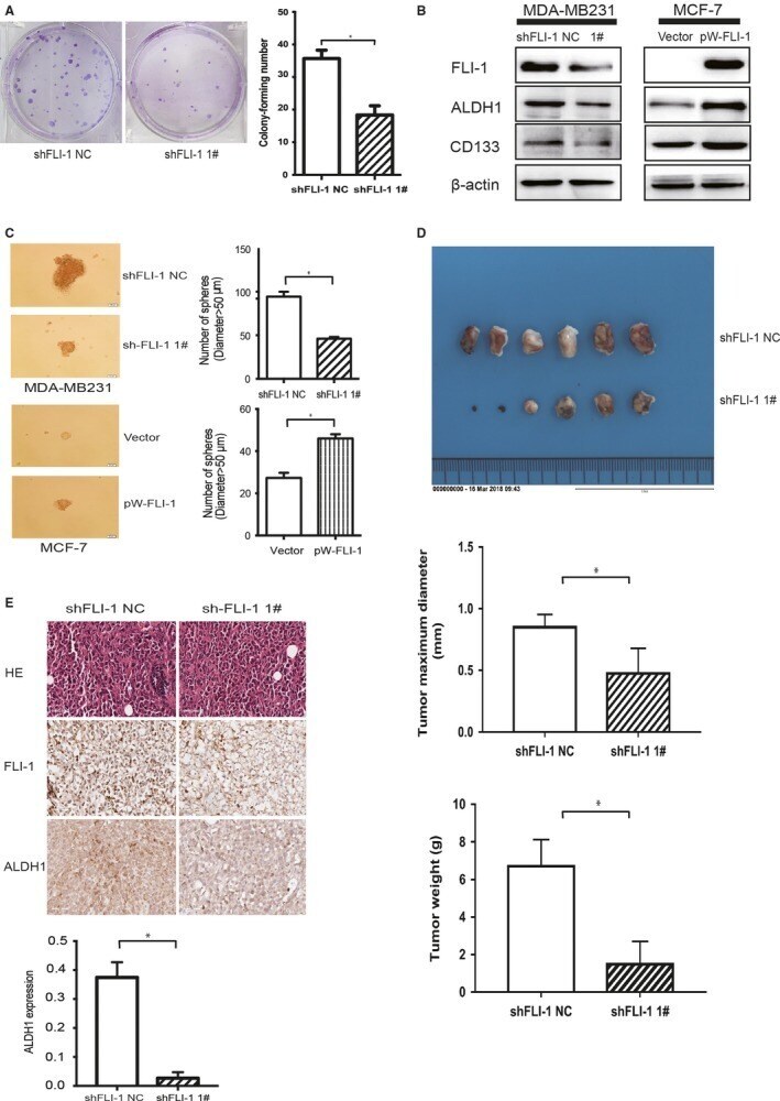
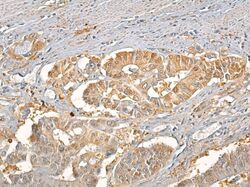
- Figure 11 Moderately differentiated adenocarcinoma with intense tumor cell reaction to ALDH1A1. Immunolabeling with anti-ALDH1A1 antibody, x200. ALDH1A1: Aldehyde dehydrogenase 1 family member A1