Explore
 Validate
 Learn
Learn Western blot
 Immunoprecipitation
Antibody data
- Antibody Data
- Antigen structure
- References [2]
- Comments [0]
- Validations
- Western blot [2]
- Immunocytochemistry [1]
- Immunohistochemistry [1]
Submit
Validation data
Reference
Comment
Report error
- Product number
- AF5869 - Provider product page

- Provider
- R&D Systems
- Product name
- Human/Mouse Aldehyde Dehydrogenase 1-A1/ ALDH1A1 Antibody
- Antibody type
- Polyclonal
- Description
- Immunogen affinity purified. Detects human Aldehyde Dehydrogenase 1-A1/ALDH1A1 in direct ELISAs and detects human and mouse Aldehyde Dehydrogenase 1-A1/ALDH1A1 in Western blots.
- Reactivity
- Human, Mouse
- Host
- Goat
- Conjugate
- Unconjugated
- Antigen sequence
P00352- Isotype
- IgG
- Vial size
- 100 ug
- Concentration
- LYOPH
- Storage
- Use a manual defrost freezer and avoid repeated freeze-thaw cycles. 12 months from date of receipt, -20 to -70 °C as supplied. 1 month, 2 to 8 °C under sterile conditions after reconstitution. 6 months, -20 to -70 °C under sterile conditions after reconstitution.
Submitted references PMP22 Regulates Self-Renewal and Chemoresistance of Gastric Cancer Cells.
Aldehyde dehydrogenase 1-positive nigrostriatal dopaminergic fibers exhibit distinct projection pattern and dopamine release dynamics at mouse dorsal striatum.
Cai W, Chen G, Luo Q, Liu J, Guo X, Zhang T, Ma F, Yuan L, Li B, Cai J
Molecular cancer therapeutics 2017 Jun;16(6):1187-1198
Molecular cancer therapeutics 2017 Jun;16(6):1187-1198
Aldehyde dehydrogenase 1-positive nigrostriatal dopaminergic fibers exhibit distinct projection pattern and dopamine release dynamics at mouse dorsal striatum.
Sgobio C, Wu J, Zheng W, Chen X, Pan J, Salinas AG, Davis MI, Lovinger DM, Cai H
Scientific reports 2017 Jul 13;7(1):5283
Scientific reports 2017 Jul 13;7(1):5283
No comments: Submit comment
Supportive validation
- Submitted by
- R&D Systems (provider)
- Main image
- Experimental details
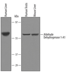
- Detection of Human and Mouse Aldehyde Dehydrogenase 1-A1/ALDH1A1 by Western Blot. Western blot shows lysates of human liver tissue, human testis tissue, and mouse liver. PVDF Membrane was probed with 1 µg/mL of Goat Anti-Human/Mouse Aldehyde Dehydrogenase 1-A1/ALDH1A1 Antigen Affinity-purified Polyclonal Antibody (Catalog # AF5869) followed by HRP-conjugated Anti-Goat IgG Secondary Antibody (Catalog # HAF019). A specific band was detected for Aldehyde Dehydrogenase 1-A1/ALDH1A1 at approximately 56 kDa (as indicated). This experiment was conducted under reducing conditions and using Immunoblot Buffer Group 8.

- Submitted by
- R&D Systems (provider)
- Main image
- Experimental details
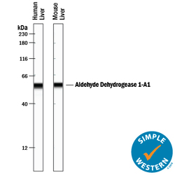
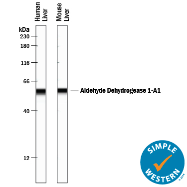
- Detection of Human and Mouse Aldehyde Dehydrogenase 1-A1/ALDH1A1 by Simple WesternTM. Simple Western lane view shows lysates of human liver tissue and mouse liver tissue, loaded at 0.2 mg/mL. A specific band was detected for Aldehyde Dehydrogenase 1-A1/ALDH1A1 at approximately 57 kDa (as indicated) using 10 µg/mL of Goat Anti-Human/Mouse Aldehyde Dehydrogenase 1-A1/ALDH1A1 Antigen Affinity-purified Polyclonal Antibody (Catalog # AF5869) followed by 1:50 dilution of HRP-conjugated Anti-Goat IgG Secondary Antibody (Catalog # HAF109). This experiment was conducted under reducing conditions and using the 12-230 kDa separation system.
Supportive validation
- Submitted by
- R&D Systems (provider)
- Main image
- Experimental details
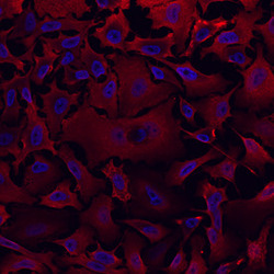
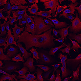
- Aldehyde Dehydrogenase 1-A1/ALDH1A1 in HeLa Human Cell Line. Aldehyde Dehydrogenase 1-A1/ALDH1A1 was detected in immersion fixed HeLa human cervical epithelial carcinoma cell line using Goat Anti-Human/Mouse Aldehyde Dehydrogenase 1-A1/ALDH1A1 Antigen Affinity-purified Polyclonal Antibody (Catalog # AF5869) at 10 µg/mL for 3 hours at room temperature. Cells were stained using the NorthernLights™ 557-conjugated Anti-Goat IgG Secondary Antibody (red; Catalog # NL001) and counterstained with DAPI (blue). Specific staining was localized to cell surfaces and cytoplasm. View our protocol for Fluorescent ICC Staining of Cells on Coverslips.
Supportive validation
- Submitted by
- R&D Systems (provider)
- Main image

- Experimental details
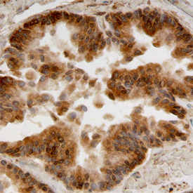
- Aldehyde Dehydrogenase 1-A1/ALDH1A1 in Human Prostate Cancer Tissue. Aldehyde Dehydrogenase 1-A1/ALDH1A1 was detected in immersion fixed paraffin-embedded sections of human prostate cancer tissue using Goat Anti-Human/Mouse Aldehyde Dehydrogenase 1-A1/ALDH1A1 Antigen Affinity-purified Polyclonal Antibody (Catalog # AF5869) at 10 µg/mL overnight at 4 °C. Before incubation with the primary antibody, tissue was subjected to heat-induced epitope retrieval using Antigen Retrieval Reagent-Basic (Catalog # CTS013). Tissue was stained using the Anti-Goat HRP-DAB Cell & Tissue Staining Kit (brown; Catalog # CTS008) and counterstained with hematoxylin (blue). Specific staining was localized to the cytoplasm of glandular epithelial cells. View our protocol for Chromogenic IHC Staining of Paraffin-embedded Tissue Sections.