 Explore
 Validate
 Learn
Learn Western blot
Antibody data
- Antibody Data
- Antigen structure
- References [0]
- Comments [0]
- Validations
- Western blot [2]
- Immunohistochemistry [6]
Submit
Validation data
Reference
Comment
Report error
- Product number
- NBP2-27406 - Provider product page

- Provider
- Novus Biologicals
- Product name
- Rat Monoclonal LIF Antibody
- Antibody type
- Monoclonal
- Description
- Protein G purified.
- Reactivity
- Human, Mouse
- Host
- Rat
- Isotype
- IgG
- Vial size
- 0.1 mg
- Concentration
- 1.0 mg/ml
- Storage
- Store at 4C short term. Aliquot and store at -20C long term. Avoid freeze-thaw cycles.
No comments: Submit comment
Supportive validation
- Submitted by
- Novus Biologicals (provider)
- Main image

- Experimental details
- Western Blot: LIF Antibody (39N7D10) [NBP2-27406] - WB validation of LIF antibody (clone 39N7D10) on (A) full-length recombinant Lif protein, (B) mouse spleen lysate and (C) human spleen lysate. 3 ug/mL concentration of primary antibody, Goat anti-rat IgG HRP secondary antibody and PicoTect ECL substrate solution were used for this assay.

- Submitted by
- Novus Biologicals (provider)
- Main image

- Experimental details
- Western Blot: LIF Antibody (39N7D10) [NBP2-27406] - LIF expression in human primary T-cells and cancer cell lines: Jurkat and K562. Image from verified customer review.

Supportive validation
- Submitted by
- Novus Biologicals (provider)
- Main image

- Experimental details
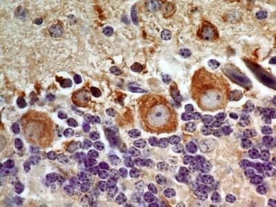
- Immunohistochemistry-Paraffin: LIF Antibody (39N7D10) [NBP2-27406] - Tissue section of adenocarcinoma of human rectum using 5 ug/ml concentration of LIF antibody (clone 39N7D10). The cancer cells as well as the goblet cells in the rectal glands depicted membrane-cytoplasmic immunostaining of LIF protein.

- Submitted by
- Novus Biologicals (provider)
- Main image

- Experimental details
- Immunohistochemistry-Paraffin: LIF Antibody (39N7D10) [NBP2-27406] - Tissue section of mouse brain using 5 ug/ml concentration of LIF antibody (clone 39N7D10).
- Submitted by
- Novus Biologicals (provider)
- Main image

- Experimental details
- Immunohistochemistry-Paraffin: LIF Antibody (39N7D10) [NBP2-27406] - Tissue section of mouse colon using 5 ug/ml concentration of LIF antibody (clone 39N7D10). The columnar epithelial cells of the crypts developed intense membrane-cytoplasmic LIF immunostaining. Additionally, some cells in the lamina propria and the sub-mucosal layer also depicted weak positivity for LIF staining.

- Submitted by
- Novus Biologicals (provider)
- Main image

- Experimental details
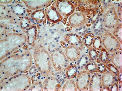
- Immunohistochemistry-Paraffin: LIF Antibody (39N7D10) [NBP2-27406] - Tissue section of mouse kidney using 5 ug/ml concentration of LIF antibody (clone 39N7D10). Very intense immune positivity of LIF was observed in membranes as well as the cytoplasm of cuboidal epithelial cells of renal tubules.

- Submitted by
- Novus Biologicals (provider)
- Main image

- Experimental details
- Immunohistochemistry-Paraffin: LIF Antibody (39N7D10) [NBP2-27406] - Tissue section of normal human kidney using 5 ug/ml concentration of LIF antibody (clone 39N7D10). Expected membrane- cytoplasmic immunepositivity of LIF was observed in the cuboidal epithelial cells of renal tubules.
- Submitted by
- Novus Biologicals (provider)
- Main image
- Experimental details
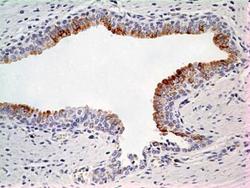
- Immunohistochemistry-Paraffin: LIF Antibody (39N7D10) [NBP2-27406] - Tissue section of normal human prostate using 5 ug/ml concentration of LIF antibody (clone 39N7D10). Cell surface/membrane- cytoplasmic immunepositivity of LIF was observed specifically in the epithelial cells of prostate alveolar glands, whereas the surrounding fibromuscular stroma cells did not develop any staining.
