Explore
 Validate
 Learn
Learn Western blot
Antibody data
- Antibody Data
- Antigen structure
- References [0]
- Comments [0]
- Validations
- Western blot [1]
- Immunohistochemistry [15]
Submit
Validation data
Reference
Comment
Report error
- Product number
- LS-C359469 - Provider product page

- Provider
- LSBio
- Product name
- LIF Antibody (aa24-203, clone 39N7D10) LS-C359469
- Antibody type
- Monoclonal
- Description
- Protein G purified
- Reactivity
- Human, Mouse
- Host
- Rat
- Isotype
- IgG
- Antibody clone number
- 39N7D10
- Storage
- Short term: store at 4°C. Long term: aliquot and store at -20°C. Avoid freeze-thaw cycles.
No comments: Submit comment
Enhanced validation
- Submitted by
- LSBio (provider)
- Enhanced method
- Genetic validation
- Main image
- Experimental details
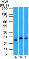
- Western Blot: LIF Antibody (39N7D10) - WB validation of LIF antibody (clone 39N7D10) on (A) full-length recombinant Lif protein, (B) mouse spleen lysate and human spleen lysate. 3 ug/ml concentration of primary antibody, Goat anti-rat Ig HRP secondary antibody and PicoTect ECL substrate solution were used for this assay. This image was taken for the unconjugated form of this product. Other forms have not been tested.
Supportive validation
- Submitted by
- LSBio (provider)
- Enhanced method
- Genetic validation
- Main image

- Experimental details
- Immunohistochemistry-Paraffin: LIF Antibody (39N7D10) - IHC analysis of formalin-fixed paraffin-embedded tissue section of mouse brain using 5 ug/ml concentration of LIF antibody (clone 39N7D10). This image was taken for the unconjugated form of this product. Other forms have not been tested.

- Submitted by
- LSBio (provider)
- Enhanced method
- Genetic validation
- Main image
- Experimental details
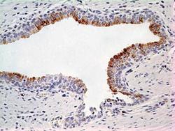
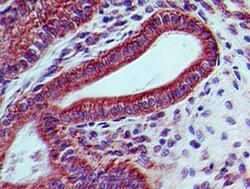
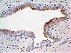
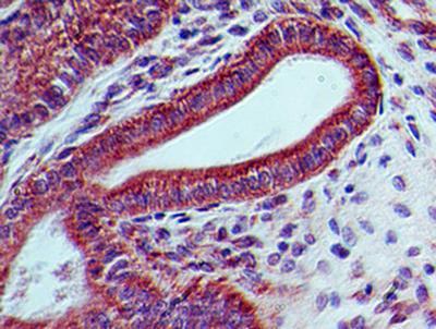
- Immunohistochemistry-Paraffin: LIF Antibody (39N7D10) - IHC analysis of formalin-fixed paraffin-embedded tissue section of normal human prostate using 5 ug/ml concentration of LIF antibody (clone 39N7D10). Cell surface/membrane- cytoplasmic immunopositivity of LIF was observed specifically in the epithelial cells of prostate alveolar glands whereas the surrounding fibromuscular stroma cells did not develop any staining. This image was taken for the unconjugated form of this product. Other forms have not been tested.

- Submitted by
- LSBio (provider)
- Enhanced method
- Genetic validation
- Main image
- Experimental details
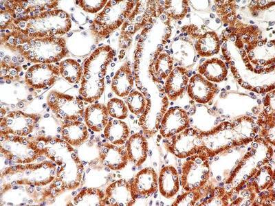
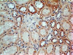
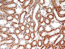
- Immunohistochemistry-Paraffin: LIF Antibody (39N7D10) - IHC analysis of formalin-fixed paraffin-embedded tissue section of normal human kidney using 5 ug/ml concentration of LIF antibody (clone 39N7D10). Expected membrane- cytoplasmic immunopositivity of LIF was observed in the cuboidal epithelial cells of renal tubules. This image was taken for the unconjugated form of this product. Other forms have not been tested.

- Submitted by
- LSBio (provider)
- Enhanced method
- Genetic validation
- Main image
- Experimental details
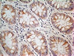
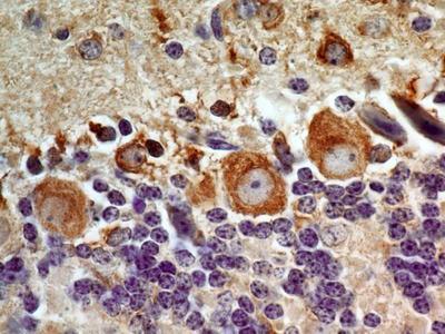
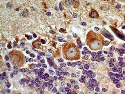
- Immunohistochemistry-Paraffin: LIF Antibody (39N7D10) - IHC analysis of formalin-fixed paraffin-embedded tissue section of adenocarcinoma of human rectum using 5 ug/ml concentration of LIF antibody (clone 39N7D10). The cancer cells as well as the goblet cells in the rectal glands depicted membrane-cytoplasmic immunostaining of LIF protein. This image was taken for the unconjugated form of this product. Other forms have not been tested.
- Submitted by
- LSBio (provider)
- Enhanced method
- Genetic validation
- Main image
- Experimental details
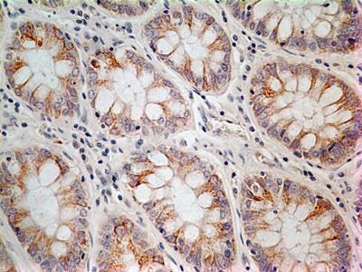
- Immunohistochemistry-Paraffin: LIF Antibody (39N7D10) - IHC analysis of formalin-fixed paraffin-embedded tissue section of mouse colon using 5 ug/ml concentration of LIF antibody (clone 39N7D10). The columnar epithelial cells of the crypts developed intense membrane-cytoplasmic LIF immunostaining. Additionally, some cells in the lamina propria and the sub-mucosal layer also depicted weak positivity for LIF staining. This image was taken for the unconjugated form of this product. Other forms have not been tested.

- Submitted by
- LSBio (provider)
- Enhanced method
- Genetic validation
- Main image
- Experimental details
- Immunohistochemistry-Paraffin: LIF Antibody (39N7D10) - IHC analysis of formalin-fixed paraffin-embedded tissue section of mouse kidney using 5 ug/ml concentration of LIF antibody (clone 39N7D10). Very intense immunopositivity of LIF was observed in membranes as well as the cytoplasm of cuboidal epithelial cells of renal tubules. This image was taken for the unconjugated form of this product. Other forms have not been tested.

- Submitted by
- LSBio (provider)
- Enhanced method
- Genetic validation
- Main image

- Experimental details
- Immunohistochemistry-Paraffin: LIF Antibody (39N7D10) - IHC analysis of formalin-fixed paraffin-embedded tissue section of mouse brain using 5 ug/ml concentration of LIF antibody (clone 39N7D10). This image was taken for the unconjugated form of this product. Other forms have not been tested.
- Submitted by
- LSBio (provider)
- Enhanced method
- Genetic validation
- Main image
- Experimental details
- Immunohistochemistry-Paraffin: LIF Antibody (39N7D10) - IHC analysis of formalin-fixed paraffin-embedded tissue section of normal human prostate using 5 ug/ml concentration of LIF antibody (clone 39N7D10). Cell surface/membrane- cytoplasmic immunopositivity of LIF was observed specifically in the epithelial cells of prostate alveolar glands whereas the surrounding fibromuscular stroma cells did not develop any staining. This image was taken for the unconjugated form of this product. Other forms have not been tested.

- Submitted by
- LSBio (provider)
- Enhanced method
- Genetic validation
- Main image

- Experimental details
- Immunohistochemistry-Paraffin: LIF Antibody (39N7D10) - IHC analysis of formalin-fixed paraffin-embedded tissue section of normal human kidney using 5 ug/ml concentration of LIF antibody (clone 39N7D10). Expected membrane- cytoplasmic immunopositivity of LIF was observed in the cuboidal epithelial cells of renal tubules. This image was taken for the unconjugated form of this product. Other forms have not been tested.

- Submitted by
- LSBio (provider)
- Enhanced method
- Genetic validation
- Main image
- Experimental details
- Immunohistochemistry-Paraffin: LIF Antibody (39N7D10) - IHC analysis of formalin-fixed paraffin-embedded tissue section of mouse brain using 5 ug/ml concentration of LIF antibody (clone 39N7D10). This image was taken for the unconjugated form of this product. Other forms have not been tested.

- Submitted by
- LSBio (provider)
- Enhanced method
- Genetic validation
- Main image

- Experimental details
- Immunohistochemistry-Paraffin: LIF Antibody (39N7D10) - IHC analysis of formalin-fixed paraffin-embedded tissue section of normal human prostate using 5 ug/ml concentration of LIF antibody (clone 39N7D10). Cell surface/membrane- cytoplasmic immunopositivity of LIF was observed specifically in the epithelial cells of prostate alveolar glands whereas the surrounding fibromuscular stroma cells did not develop any staining. This image was taken for the unconjugated form of this product. Other forms have not been tested.

- Submitted by
- LSBio (provider)
- Enhanced method
- Genetic validation
- Main image

- Experimental details
- Immunohistochemistry-Paraffin: LIF Antibody (39N7D10) - IHC analysis of formalin-fixed paraffin-embedded tissue section of normal human kidney using 5 ug/ml concentration of LIF antibody (clone 39N7D10). Expected membrane- cytoplasmic immunopositivity of LIF was observed in the cuboidal epithelial cells of renal tubules. This image was taken for the unconjugated form of this product. Other forms have not been tested.

- Submitted by
- LSBio (provider)
- Enhanced method
- Genetic validation
- Main image

- Experimental details
- Immunohistochemistry-Paraffin: LIF Antibody (39N7D10) - IHC analysis of formalin-fixed paraffin-embedded tissue section of adenocarcinoma of human rectum using 5 ug/ml concentration of LIF antibody (clone 39N7D10). The cancer cells as well as the goblet cells in the rectal glands depicted membrane-cytoplasmic immunostaining of LIF protein. This image was taken for the unconjugated form of this product. Other forms have not been tested.

- Submitted by
- LSBio (provider)
- Enhanced method
- Genetic validation
- Main image

- Experimental details
- Immunohistochemistry-Paraffin: LIF Antibody (39N7D10) - IHC analysis of formalin-fixed paraffin-embedded tissue section of mouse colon using 5 ug/ml concentration of LIF antibody (clone 39N7D10). The columnar epithelial cells of the crypts developed intense membrane-cytoplasmic LIF immunostaining. Additionally, some cells in the lamina propria and the sub-mucosal layer also depicted weak positivity for LIF staining. This image was taken for the unconjugated form of this product. Other forms have not been tested.
- Submitted by
- LSBio (provider)
- Enhanced method
- Genetic validation
- Main image

- Experimental details
- Immunohistochemistry-Paraffin: LIF Antibody (39N7D10) - IHC analysis of formalin-fixed paraffin-embedded tissue section of mouse kidney using 5 ug/ml concentration of LIF antibody (clone 39N7D10). Very intense immunopositivity of LIF was observed in membranes as well as the cytoplasm of cuboidal epithelial cells of renal tubules. This image was taken for the unconjugated form of this product. Other forms have not been tested.