Explore
 Validate
 Learn
Learn Immunohistochemistry
 Blocking/Neutralizing
Antibody data
- Antibody Data
- Antigen structure
- References [0]
- Comments [0]
- Validations
- Immunohistochemistry [2]
- Other assay [2]
Submit
Validation data
Reference
Comment
Report error
- Product number
- PA5-47337 - Provider product page

- Provider
- Invitrogen Antibodies
- Product name
- LIF Polyclonal Antibody
- Antibody type
- Polyclonal
- Antigen
- Recombinant full-length protein
- Description
- In direct ELISAs and Western blots, less than 30% cross-reactivity with recombinant mouse LIF is observed. Reconstitute at 0.2 mg/mL in sterile PBS. Endoxin level is
- Reactivity
- Human
- Host
- Goat
- Isotype
- IgG
- Vial size
- 100 μg
- Concentration
- 0.2 mg/mL
- Storage
- -20°C, Avoid Freeze/Thaw Cycles
No comments: Submit comment
Supportive validation
- Submitted by
- Invitrogen Antibodies (provider)
- Main image
- Experimental details
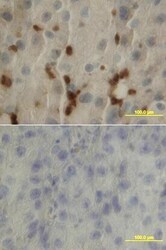
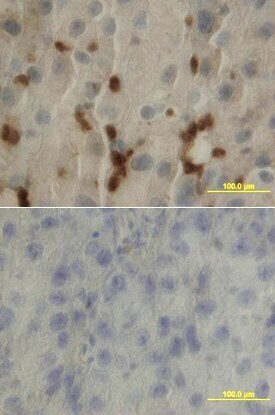
- Immunohistochemical analysis of LIF in immersion fixed paraffin-embedded sections of human lung array. Samples were incubated in LIF polyclonal antibody (Product # PA5-47337) using a dilution of 25 µg/mL overnight at 4 °C. Tissue was stained using the Anti-Goat HRP-DAB Cell & Tissue Staining Kit (brown) and counterstained with hematoxylin (blue). Lower panel shows a lack of labeling if primary antibodies are omitted and tissue is stained only with secondary antibody followed by incubation with detection reagents.
- Submitted by
- Invitrogen Antibodies (provider)
- Main image

- Experimental details
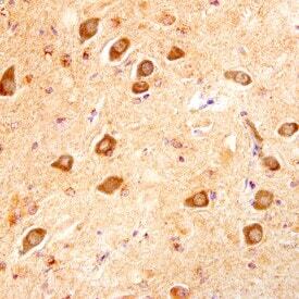
- Immunohistochemical analysis of LIF in immersion fixed paraffin-embedded sections of human Alzheimers brain. Samples were incubated in LIF polyclonal antibody (Product # PA5-47337) using a dilution of 10 µg/mL overnight at 4 °C. Before incubation with the primary antibody, tissue was subjected to heat-induced epitope retrieval using Antigen Retrieval Reagent-Basic . Tissue was stained using the Anti-Goat HRP-DAB Cell & Tissue Staining Kit (brown) and counterstained with hematoxylin (blue). Specific staining was localized to neuronal cytoplasm.
Supportive validation
- Submitted by
- Invitrogen Antibodies (provider)
- Main image
- Experimental details
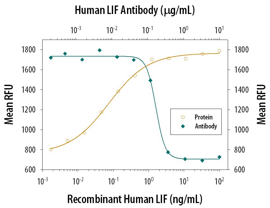
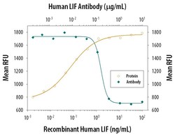
- Neutralization of LIF in TF‚1 human erythroleukemic cell line. Samples were incubated in LIF polyclonal antibody (Product # PA5-47337). Recombinant Human LIF stimulates proliferation in the TF‚1 human erythroleukemic cell line in a dose-dependent manner (orange line), as measured by Resazurin. Proliferation elicited by 3 ng/mL Recombinant Human LIF is neutralized (green line) by increasing concentrations of Goat Anti-Human LIF Antigen Affinity-purified Polyclonal Antibody. The ND50 is typically 0.04‚0.08 µg/mL.

- Submitted by
- Invitrogen Antibodies (provider)
- Main image

- Experimental details
- Neutralization of LIF in TF‚1 human erythroleukemic cell line. Samples were incubated in LIF polyclonal antibody (Product # PA5-47337). Recombinant Human LIF stimulates proliferation in the TF‚1 human erythroleukemic cell line in a dose-dependent manner (orange line), as measured by Resazurin. Proliferation elicited by 3 ng/mL Recombinant Human LIF is neutralized (green line) by increasing concentrations of Goat Anti-Human LIF Antigen Affinity-purified Polyclonal Antibody. The ND50 is typically 0.04‚0.08 µg/mL.