Explore
 Validate
 Learn
Learn Western blot
Antibody data
- Antibody Data
- Antigen structure
- References [0]
- Comments [0]
- Validations
- Western blot [2]
- Flow cytometry [1]
Submit
Validation data
Reference
Comment
Report error
- Product number
- MAB7966 - Provider product page

- Provider
- Novus Biologicals
- Product name
- Mouse Monoclonal TMEM87A Antibody
- Antibody type
- Monoclonal
- Description
- Protein A or G purified from hybridoma culture supernatant. Detects human TMEM87A in ELISAs and Western Blots
- Reactivity
- Human
- Host
- Mouse
- Conjugate
- Unconjugated
- Isotype
- IgG
- Vial size
- 100 ug
- Concentration
- LYOPH
- Storage
- Use a manual defrost freezer and avoid repeated freeze-thaw cycles. 12 months from date of receipt, -20 to -70 degreesC as supplied. 1 month, 2 to 8 degreesC under sterile conditions after reconstitution. 6 months, -20 to -70 degreesC under sterile conditions after reconstitution.
No comments: Submit comment
Supportive validation
- Submitted by
- Novus Biologicals (provider)
- Main image

- Experimental details
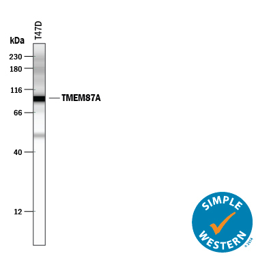
- Detection of Human TMEM87A by Simple WesternTM. Simple Western lane view shows lysate of T47D human breast cancer cell line, loaded at 0.5 mg/mL. Specific bands were detected for TMEM87A at approximately 98 kDa (as indicated) using 2 µg/mL of Mouse Anti-Human TMEM87A Monoclonal Antibody (Catalog # MAB7966). This experiment was conducted under reducing conditions and using the 12-230 kDa separation system.
- Submitted by
- Novus Biologicals (provider)
- Main image
- Experimental details
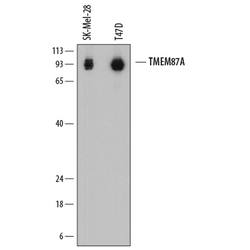
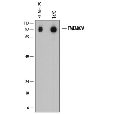
- Detection of Human TMEM87A by Western Blot. Western blot shows lysates of SK-Mel-28 human malignant melanoma cell line and T47D human breast cancer cell line. PVDF membrane was probed with 0.2 µg/mL of Mouse Anti-Human TMEM87A Monoclonal Antibody (Catalog # MAB7966) followed by HRP-conjugated Anti-Mouse IgG Secondary Antibody (Catalog # HAF018). Specific bands were detected for TMEM87A at approximately 90-95 kDa (as indicated). This experiment was conducted under reducing conditions and using Immunoblot Buffer Group 1.
Supportive validation
- Submitted by
- Novus Biologicals (provider)
- Main image

- Experimental details
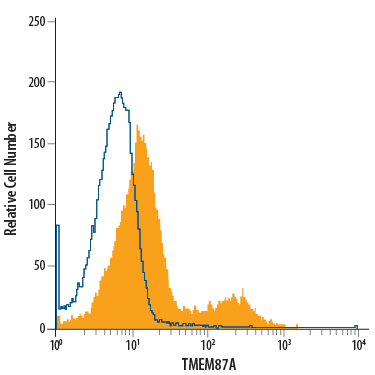
- Detection of TMEM-87A in PC-3 Human Cell Line by Flow Cytometry. PC-3 human prostate cancer cell line was stained with Mouse Anti-Human TMEM87A Monoclonal Antibody (Catalog # MAB7966, filled histogram) or isotype control antibody (Catalog # MAB0041, open histogram), followed by Allophycocyanin-conjugated Anti-Mouse IgG Secondary Antibody (Catalog # F0101B).