Explore
 Validate
 Learn
Learn Western blot
 Immunohistochemistry
Antibody data
- Antibody Data
- Antigen structure
- References [8]
- Comments [0]
- Validations
- Immunohistochemistry [2]
- Blocking/Neutralizing [1]
Submit
Validation data
Reference
Comment
Report error
- Product number
- AF-495-NA - Provider product page

- Provider
- R&D Systems
- Product name
- Mouse Oncostatin M/OSM Antibody
- Antibody type
- Polyclonal
- Description
- Antigen Affinity-purified. Detects mouse OSM in direct ELISAs and Western blots. In direct ELISAs, less than 2% cross-reactivity with recombinant human OSM is observed.
- Reactivity
- Mouse
- Host
- Goat
- Conjugate
- Unconjugated
- Antigen sequence
P53347- Isotype
- IgG
- Vial size
- 100 ug
- Concentration
- LYOPH
- Storage
- Use a manual defrost freezer and avoid repeated freeze-thaw cycles. 12 months from date of receipt, -20 to -70 °C as supplied. 1 month, 2 to 8 °C under sterile conditions after reconstitution. 6 months, -20 to -70 °C under sterile conditions after reconstitution.
Submitted references Neutrophil extracellular traps (NETs) contribute to pathological changes of ocular graft-vs.-host disease (oGVHD) dry eye: Implications for novel biomarkers and therapeutic strategies.
Anti-OSM Antibody Inhibits Tubulointerstitial Lesion in a Murine Model of Lupus Nephritis.
Oncostatin M overexpression induces skin inflammation but is not required in the mouse model of imiquimod-induced psoriasis-like inflammation.
Oncostatin m, an inflammatory cytokine produced by macrophages, supports intramembranous bone healing in a mouse model of tibia injury.
Oncostatin M acting via OSMR, augments the actions of IL-1 and TNF in synovial fibroblasts.
Activation of NFAT signaling establishes a tumorigenic microenvironment through cell autonomous and non-cell autonomous mechanisms.
Long term oncostatin M treatment induces an osteocyte-like differentiation on osteosarcoma and calvaria cells.
Cardiotrophin-1 in choroid plexus and the cerebrospinal fluid circulatory system.
An S, Raju I, Surenkhuu B, Kwon JE, Gulati S, Karaman M, Pradeep A, Sinha S, Mun C, Jain S
The ocular surface 2019 Jul;17(3):589-614
The ocular surface 2019 Jul;17(3):589-614
Anti-OSM Antibody Inhibits Tubulointerstitial Lesion in a Murine Model of Lupus Nephritis.
Liu Q, Du Y, Li K, Zhang W, Feng X, Hao J, Li H, Liu S
Mediators of inflammation 2017;2017:3038514
Mediators of inflammation 2017;2017:3038514
Oncostatin M overexpression induces skin inflammation but is not required in the mouse model of imiquimod-induced psoriasis-like inflammation.
Pohin M, Guesdon W, Mekouo AA, Rabeony H, Paris I, Atanassov H, Favot L, Mcheik J, Bernard FX, Richards CD, Amiaud J, Blanchard F, Lecron JC, Morel F, Jégou JF
European journal of immunology 2016 Jul;46(7):1737-51
European journal of immunology 2016 Jul;46(7):1737-51
Oncostatin m, an inflammatory cytokine produced by macrophages, supports intramembranous bone healing in a mouse model of tibia injury.
Guihard P, Boutet MA, Brounais-Le Royer B, Gamblin AL, Amiaud J, Renaud A, Berreur M, Rédini F, Heymann D, Layrolle P, Blanchard F
The American journal of pathology 2015 Mar;185(3):765-75
The American journal of pathology 2015 Mar;185(3):765-75
Oncostatin M acting via OSMR, augments the actions of IL-1 and TNF in synovial fibroblasts.
Le Goff B, Singbrant S, Tonkin BA, Martin TJ, Romas E, Sims NA, Walsh NC
Cytokine 2014 Aug;68(2):101-9
Cytokine 2014 Aug;68(2):101-9
Activation of NFAT signaling establishes a tumorigenic microenvironment through cell autonomous and non-cell autonomous mechanisms.
Tripathi P, Wang Y, Coussens M, Manda KR, Casey AM, Lin C, Poyo E, Pfeifer JD, Basappa N, Bates CM, Ma L, Zhang H, Pan M, Ding L, Chen F
Oncogene 2014 Apr 3;33(14):1840-9
Oncogene 2014 Apr 3;33(14):1840-9
Long term oncostatin M treatment induces an osteocyte-like differentiation on osteosarcoma and calvaria cells.
Brounais B, David E, Chipoy C, Trichet V, Ferré V, Charrier C, Duplomb L, Berreur M, Rédini F, Heymann D, Blanchard F
Bone 2009 May;44(5):830-9
Bone 2009 May;44(5):830-9
Cardiotrophin-1 in choroid plexus and the cerebrospinal fluid circulatory system.
Gard AL, Gavin E, Solodushko V, Pennica D
Neuroscience 2004;127(1):43-52
Neuroscience 2004;127(1):43-52
No comments: Submit comment
Supportive validation
- Submitted by
- R&D Systems (provider)
- Main image
- Experimental details
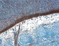
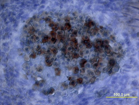
- Oncostatin M/OSM in Mouse Embryo. Oncostatin M/OSM was detected in immersion fixed frozen sections of mouse embryo (15 d.p.c, section through spinal cord) using Mouse Oncostatin M/OSM Antigen Affinity-purified Polyclonal Antibody (Catalog # AF-495-NA) at 15 µg/mL overnight at 4 °C. Tissue was stained using the Anti-Goat HRP-DAB Cell & Tissue Staining Kit (brown; Catalog # CTS008) and counterstained with hematoxylin (blue). View our protocol for Chromogenic IHC Staining of Frozen Tissue Sections.
- Submitted by
- R&D Systems (provider)
- Main image
- Experimental details
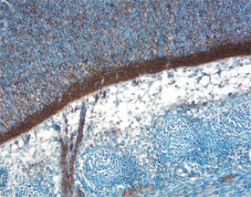
- Oncostatin M/OSM in Mouse Embryo. Oncostatin M/OSM was detected in immersion fixed frozen sections of mouse embryo using Mouse Oncostatin M/OSM Antigen Affinity-purified Polyclonal Antibody (Catalog # AF-495-NA) at 15 µg/mL overnight at 4 °C. Tissue was stained using the Anti-Goat HRP-DAB Cell & Tissue Staining Kit (brown; Catalog # CTS008) and counterstained with hematoxylin (blue). View our protocol for Chromogenic IHC Staining of Frozen Tissue Sections.
Supportive validation
- Submitted by
- R&D Systems (provider)
- Main image
- Experimental details
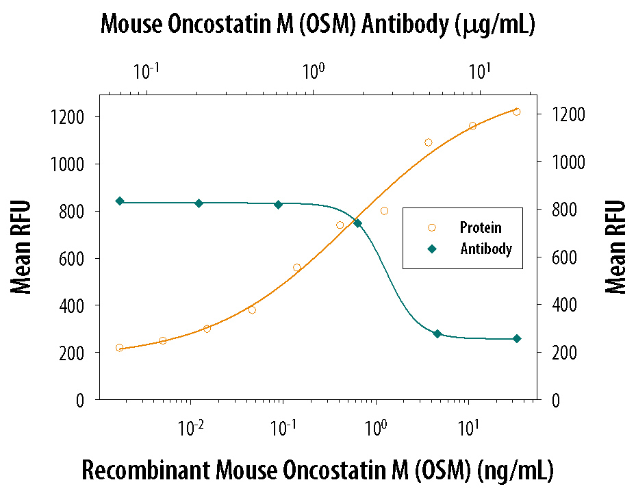
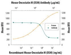
- Cell Proliferation Induced by Oncostatin M/OSM and Neutralization by Mouse Oncostatin M/OSM Antibody. Recombinant Mouse Oncostatin M/OSM (Catalog # 495-MO) stimulates proliferation in the NIH-3T3 mouse embryonic fibroblast cell line in a dose-dependent manner (orange line). Proliferation elicited by Recombinant Mouse Oncostatin M/OSM (15 ng/mL) is neutralized (green line) by increasing concentrations of Mouse Oncostatin M/OSM Antigen Affinity-purified Polyclonal Antibody (Catalog # AF-495-NA). The ND50 is typically 0.6-3.0 µg/mL.