 Explore
 Validate
 Learn
Learn Western blot
 Other assay
Antibody data
- Antibody Data
- Antigen structure
- References [1]
- Comments [0]
- Validations
- Other assay [1]
Submit
Validation data
Reference
Comment
Report error
- Product number
- PA5-23298 - Provider product page

- Provider
- Invitrogen Antibodies
- Product name
- RAP2A Polyclonal Antibody
- Antibody type
- Polyclonal
- Antigen
- Other
- Reactivity
- Human, Mouse, Canine, Chicken/Avian, Xenopus
- Host
- Rabbit
- Isotype
- IgG
- Vial size
- 100 μg
- Concentration
- 1.0 mg/mL
- Storage
- -20°C, Avoid Freeze/Thaw Cycles
Submitted references Data in support of Rap2a GTPase expression, activation and effects in LPS-mediated innate immune response and NF-κB activation.
Carvalho BC, Oliveira LC, Rocha CD, Fernandes HB, Oliveira IM, Leãõ FB, Valverde TM, Rego IMG, Ghosh S, Silva AM
Data in brief 2019 Jun;24:103965
Data in brief 2019 Jun;24:103965
No comments: Submit comment
Supportive validation
- Submitted by
- Invitrogen Antibodies (provider)
- Main image
- Experimental details
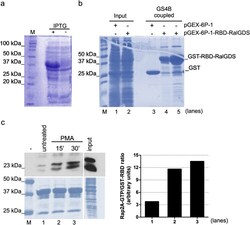
- Fig. 2 Expression of GST-RBD-RalGDS and validation of affinity precipitation of Rap2a in mammalian cell extracts. ( a ) The expression of GST-RBD-RalGDS was obtained upon induction with 1 mM isopropyl-1-thio-beta- d -galactopyranoside (IPTG) for 2 hours of bacterial cell cultures transformed with pGEX-6P-1-RBD-RalGDS. Bacterial extracts were fractionated onto 10% SDS-PAGE, and followed by coomassie blue staining. ( b ) Coupling of GST-RBD-RalGDS to glutathione sepharose 4B (GS4B) beads. Suspensions of IPTG-induced bacterial cell cultures transformed with pGEX-6P-1 or pGEX-6P-1-RBD-RalGDS were centrifuged, lysed and sonicated. GST or GST-RBD-RalGDS fusion protein were mixed with GS4B. Bacterial cell lysates (lanes 1 and 2) and eluted samples (lanes 3-5) were separated by 10% SDS-PAGE, followed by coomassie blue staining. Two independent bacterial clones of pGEX-6P-1-RBD-RalGDS were used in lanes 4 and 5, respectively. ( c ) RAW264 cells were treated with PMA (100nM) as indicated. The lysates (500 mug) were then incubated with 100mul (~0.5mg) of bacterial lysates containing GST-RalGDS-RBD precoupled to GS4B beads. After washes, the beads mixtures were fractionated on 12% SDS-PAGE, transferred to PVDF membrane, and probed with anti-Rap2A antibody. Anti-Rabbit IgG (H + L), peroxidase conjugated was used as the secondary antibody. The detection was performed with Clarity Western ECL Blotting Substrate (BioRad) and followed by exposure to X-ray film. Densitometrical analysis of the
