 Explore
 Validate
 Learn
Learn Western blot
 Chromatin Immunoprecipitation
Antibody data
- Antibody Data
- Antigen structure
- References [0]
- Comments [0]
- Validations
- Western blot [4]
- Immunocytochemistry [3]
- Immunohistochemistry [1]
Submit
Validation data
Reference
Comment
Report error
- Product number
- GTX102835 - Provider product page

- Provider
- GeneTex
- Proper citation
- GeneTex Cat#GTX102835, RRID:AB_11164652
- Product name
- AF9 antibody
- Antibody type
- Polyclonal
- Reactivity
- Human
- Host
- Rabbit
No comments: Submit comment
Supportive validation
- Submitted by
- GeneTex (provider)
- Main image

- Experimental details
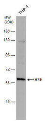
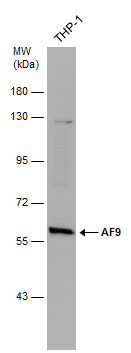
- Sample (30 ?g of whole cell lysate) A: THP-1 7.5% SDS PAGE GTX102835 diluted at 1:1000 The HRP-conjugated anti-rabbit IgG antibody (GTX213110-01) was used to detect the primary antibody.
- Submitted by
- GeneTex (provider)
- Main image
- Experimental details
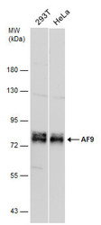
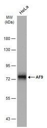
- Various whole cell extracts (30 ?g) were separated by 7.5% SDS-PAGE, and the membrane was blotted with AF9 antibody (GTX102835) diluted at 1:1000.
- Submitted by
- GeneTex (provider)
- Main image
- Experimental details
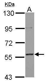
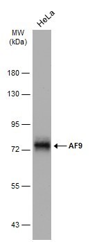
- Whole cell extract (30 ?g) was separated by 7.5% SDS-PAGE, and the membrane was blotted with AF9 antibody (GTX102835) diluted at 1:500.
- Submitted by
- GeneTex (provider)
- Main image
- Experimental details
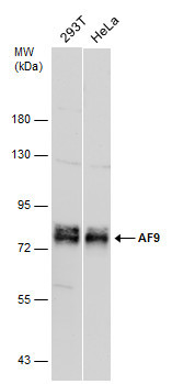
- Whole cell extract (30 ?g) was separated by 7.5% SDS-PAGE, and the membrane was blotted with AF9 antibody (GTX102835) diluted at 1:1000.
Supportive validation
- Submitted by
- GeneTex (provider)
- Main image
- Experimental details
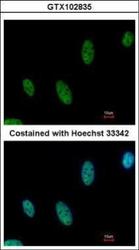
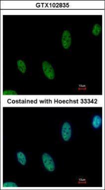
- Immunofluorescence analysis of paraformaldehyde-fixed HeLa, using AF9(GTX102835) antibody at 1:500 dilution.
- Submitted by
- GeneTex (provider)
- Main image

- Experimental details
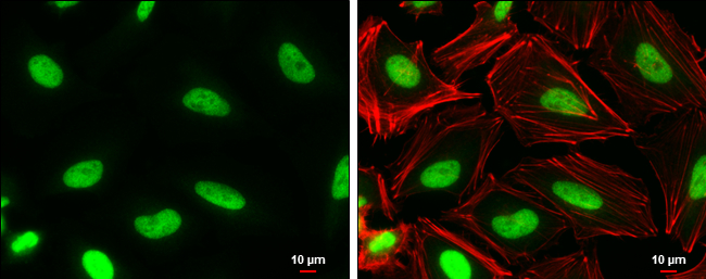
- AF9 antibody detects AF9 protein at nucleus by immunofluorescent analysis.Sample: HeLa cells were fixed in 4% paraformaldehyde at RT for 15 min.Green: AF9 protein stained by AF9 antibody (GTX102835) diluted at 1:1000.Red: Phalloidin, a cytoskeleton marker, diluted at 1:100.Scale bar = 10 £gm.
- Submitted by
- GeneTex (provider)
- Main image

- Experimental details
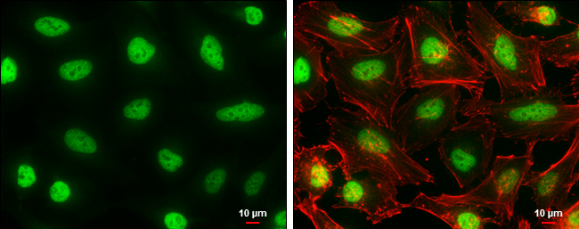
- AF9 antibody detects AF9 protein at nucleus by immunofluorescent analysis.Sample: HeLa cells were fixed in 4% paraformaldehyde at RT for 15 min.Green: AF9 protein stained by AF9 antibody (GTX102835) diluted at 1:1000.Red: Phalloidin, a cytoskeleton marker, diluted at 1:100.Scale bar = 10 £gm.
Supportive validation
- Submitted by
- GeneTex (provider)
- Main image
- Experimental details
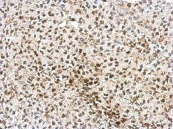
- Immunohistochemical analysis of paraffin-embedded D54 xenograft, using AF9(GTX102835) antibody at 1:750 dilution.
