Explore
 Validate
 Learn
LearnALX-804-366-C100
antibody from Enzo Life Sciences
Targeting: DIABLO
DFNA64, DIABLO-S, FLJ10537, FLJ25049, SMAC
 Western blot
 ELISA
ELISA Immunocytochemistry
Antibody data
- Antibody Data
- Antigen structure
- References [3]
- Comments [0]
- Validations
- Immunocytochemistry [3]
Submit
Validation data
Reference
Comment
Report error
- Product number
- ALX-804-366-C100 - Provider product page

- Provider
- Enzo Life Sciences
- Proper citation
- Enzo Life Sciences Cat#ALX-804-366-C100, RRID:AB_2052567
- Product name
- Smac/DIABLO (mouse) monoclonal antibody (9H10)
- Antibody type
- Monoclonal
- Antigen
- Recombinant protein fragment
- Reactivity
- Mouse
- Host
- Rat
- Isotype
- IgG
- Antibody clone number
- 9H10
- Vial size
- 100 μg
- Storage
- -20°C
- Handling
- Avoid freeze/thaw cycles.
Submitted references Treatment of mice with 2,3,7,8-tetrachlorodibenzo-p-dioxin leads to aryl hydrocarbon receptor-dependent nuclear translocation of NF-kappaB and expression of Fas ligand in thymic stromal cells and consequent apoptosis in T cells.
Tissue distribution of Diablo/Smac revealed by monoclonal antibodies.
Bcl-rambo, a novel Bcl-2 homologue that induces apoptosis via its unique C-terminal extension.
Camacho IA, Singh N, Hegde VL, Nagarkatti M, Nagarkatti PS
Journal of immunology (Baltimore, Md. : 1950) 2005 Jul 1;175(1):90-103
Journal of immunology (Baltimore, Md. : 1950) 2005 Jul 1;175(1):90-103
Tissue distribution of Diablo/Smac revealed by monoclonal antibodies.
Tikoo A, O'Reilly L, Day CL, Verhagen AM, Pakusch M, Vaux DL
Cell death and differentiation 2002 Jul;9(7):710-6
Cell death and differentiation 2002 Jul;9(7):710-6
Bcl-rambo, a novel Bcl-2 homologue that induces apoptosis via its unique C-terminal extension.
Kataoka T, Holler N, Micheau O, Martinon F, Tinel A, Hofmann K, Tschopp J
The Journal of biological chemistry 2001 Jun 1;276(22):19548-54
The Journal of biological chemistry 2001 Jun 1;276(22):19548-54
No comments: Submit comment
Supportive validation
- Submitted by
- Enzo Life Sciences (provider)
- Main image
- Experimental details
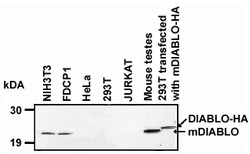
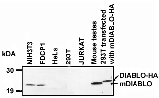
- Detection of endogenous and over-expressed DIABLO by Western blot by clone 9H10. When analysis of endogenous DIABLO (50 to 100µg total protein per lane) in healthy cells of human as well as mouse origin is performed, only the processed form of mouse DIABLO (NIH3T3 and FDCP1 mouse cell lines) can be detected. The processed form of mouse DIABLO is detectable when over-expressed in human 293T cells serving as a positive control. The HA-tagged mDIABLO migrates at a slightly higher molecular weight than endogenous wild type mouse DIABLO (mouse testes). Method: Recommended dilution for MAb to Smac/DIABLO (clone 9H10): 1: 500 to 1:1'000 for 4-6 h at room temperature or 4¡C o/n, followed by incubation with an anti-rat IgG HRP-conjugated antibody (Prod. No. ALX-211-052) at a dilution 1:2'000 to 1:5'000. Exposure time was 10-30 mins (Pico chemoluminiscence detection system Pierce). Technical note: Only secondary anti-rat IgG antibodies of the highest quality are recommended for use with 9H10 in Western blot (PAb to Rat IgG (Mouse Ig adsorbed) (HRP), Prod. No.ALX-211-052) and ICC (PAb to Rat IgG (Mouse Ig adsorbed) (FITC), Prod. No. ALX-211-051).
- Submitted by
- Enzo Life Sciences (provider)
- Main image

- Experimental details
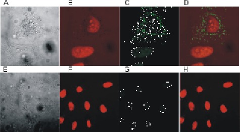
- Mouse Smac/DIABLO is associated with the mitochondria when overexpressed in human HeLa cells, nuclei are counterstained with propidium iodide (PI). Method: MAb to Smac/DIABLO (clone 9H10) was used at a dilution of 1:100 (in C, phase contrast in A, staining with PI in B, overlay with PI shown in D), but the optimal concentration needs to be determined individually. Cells were fixed with 4% PFA and permeabilized with 0.1-0.3% saponin, followed by incubation with an anti-rat-ALEXA490 conjugated antibody (Molecular Probes). Staining with a negative control (rat IgG) is shown in E to H.
- Submitted by
- Enzo Life Sciences (provider)
- Main image
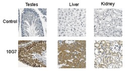
- Experimental details
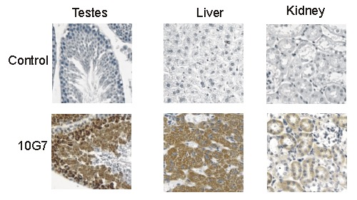
- Immunohistochemical detection of mouse Smac/DIABLO by clone 9H10. Method: Freshly cut mouse tissues were fixed, paraffin embedded and cut. Before staining the tissue sections were dewaxed and gradually rehydrated. Tissue sections were treated with 0.3% hydrogen peroxide in PBS to block the endogenous peroxidase activity. The cells were then permeabilized by incubation in 0.2% Triton X-100 and non-specific staining was prevented by incubation with 2.5% horse serum for 1 hr at room temperature. Sections were incubated in a humidified chamber with clone 9H10 at 10 µg/ml for 1 hr at room temperature. After washing in PBS containing 0.2% Triton X-100, tissues were incubated with HRP conjugated goat anti-rat IgG. To enhance the signal, the tissues were incubated with biotinylated goat anti-rat IgG (Prod. No. ALX-211-058). This was followed by incubating with ABC Vectastain Elite reagent (Prod. No. VC-PK-6100) and finally the sections were stained with DAB reagent (Prod. No. VC-SK-4100) according to the manufacturer's instructions. Sections were counterstained with hematoxylin (Prod. No. VC-H-3401), and dehydrated in graded concentrations of alcohol and histolene before mounting.