 Explore
 Validate
 Learn
Learn Western blot
Antibody data
- Antibody Data
- Antigen structure
- References [3]
- Comments [0]
- Validations
- Western blot [1]
Submit
Validation data
Reference
Comment
Report error
- Product number
- PAB16923 - Provider product page

- Provider
- Abnova Corporation
- Proper citation
- Abnova Corporation Cat#PAB16923, RRID:AB_10678030
- Product name
- IL32 polyclonal antibody
- Antibody type
- Polyclonal
- Description
- Rabbit polyclonal antibody raised against full length recombinant IL32.
- Storage
- Store at 4°C on dry atmosphere.After reconstitution with 0.1 mL of deionized water, store at -20°C or lower.Aliquot to avoid repeated freezing and thawing.
Submitted references Molecular characterization of IL-32 in human endothelial cells.
Interleukin-32 expression in the pancreas.
Negative feedback regulation of IL-32 production by iNOS activation in response to dsRNA or influenza virus infection.
Kobayashi H, Lin PC
Cytokine 2009 Jun;46(3):351-8
Cytokine 2009 Jun;46(3):351-8
Interleukin-32 expression in the pancreas.
Nishida A, Andoh A, Inatomi O, Fujiyama Y
The Journal of biological chemistry 2009 Jun 26;284(26):17868-76
The Journal of biological chemistry 2009 Jun 26;284(26):17868-76
Negative feedback regulation of IL-32 production by iNOS activation in response to dsRNA or influenza virus infection.
Li W, Yang F, Liu Y, Gong R, Liu L, Feng Y, Hu P, Sun W, Hao Q, Kang L, Wu J, Zhu Y
European journal of immunology 2009 Apr;39(4):1019-24
European journal of immunology 2009 Apr;39(4):1019-24
No comments: Submit comment
Supportive validation
- Submitted by
- Abnova Corporation (provider)
- Main image
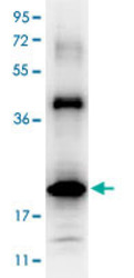
- Experimental details
- Western blot using IL32 polyclonal antibody (Cat # PAB16923) shows detection of a band ~19 KDa in size corresponding to recombinant human IL32. After transfer, the membrane was blocked overnight with 3% BSA in TBS followed by reaction with primary antibody at a 1 : 1,000 dilution. Detection occurred using peroxidase conjugated anti-Rabbit IgG secondary antibody diluted 1 : 40,000 in blocking buffer for 30 min at RT followed by reaction with FemtoMax™ chemiluminescent substrate. Image was captured using VersaDoc™ MP 4000 imaging system (Bio-Rad).
