 Explore
 Validate
 Learn
Learn Western blot
Antibody data
- Antibody Data
- Antigen structure
- References [3]
- Comments [0]
- Validations
- Western blot [1]
- Immunocytochemistry [1]
Submit
Validation data
Reference
Comment
Report error
- Product number
- MAB30401 - Provider product page

- Provider
- R&D Systems
- Product name
- Human IL-32 alpha Antibody
- Antibody type
- Monoclonal
- Description
- Protein A or G purified from hybridoma culture supernatant. Detects the alpha isoform of IL-32 in direct ELISAs. In direct ELISAs, less than 15% cross-reactivity with recombinant human (rh) IL-32 beta and rhIL-32 gamma is observed.
- Reactivity
- Human
- Host
- Rat
- Conjugate
- Unconjugated
- Antigen sequence
NP_001012651- Isotype
- IgG
- Antibody clone number
- 373802
- Vial size
- 100 ug
- Concentration
- LYOPH
- Storage
- Use a manual defrost freezer and avoid repeated freeze-thaw cycles. 12 months from date of receipt, -20 to -70 °C as supplied. 1 month, 2 to 8 °C under sterile conditions after reconstitution. 6 months, -20 to -70 °C under sterile conditions after reconstitution.
Submitted references Allele-specific long-distance regulation dictates IL-32 isoform switching and mediates susceptibility to HIV-1.
Increased Interleukin-32 Levels in Obesity Promote Adipose Tissue Inflammation and Extracellular Matrix Remodeling: Effect of Weight Loss.
Native IL-32 is released from intestinal epithelial cells via a non-classical secretory pathway as a membrane-associated protein.
Palstra RJ, de Crignis E, Röling MD, van Staveren T, Kan TW, van Ijcken W, Mueller YM, Katsikis PD, Mahmoudi T
Science advances 2018 Feb;4(2):e1701729
Science advances 2018 Feb;4(2):e1701729
Increased Interleukin-32 Levels in Obesity Promote Adipose Tissue Inflammation and Extracellular Matrix Remodeling: Effect of Weight Loss.
Catalán V, Gómez-Ambrosi J, Rodríguez A, Ramírez B, Valentí V, Moncada R, Landecho MF, Silva C, Salvador J, Frühbeck G
Diabetes 2016 Dec;65(12):3636-3648
Diabetes 2016 Dec;65(12):3636-3648
Native IL-32 is released from intestinal epithelial cells via a non-classical secretory pathway as a membrane-associated protein.
Hasegawa H, Thomas HJ, Schooley K, Born TL
Cytokine 2011 Jan;53(1):74-83
Cytokine 2011 Jan;53(1):74-83
No comments: Submit comment
Supportive validation
- Submitted by
- R&D Systems (provider)
- Main image
- Experimental details
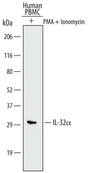
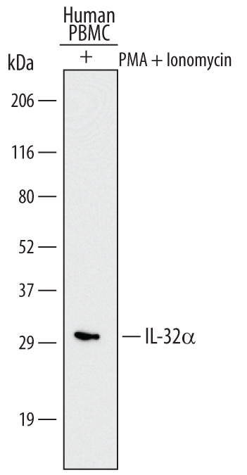
- Detection of Human IL-32 alpha by Western Blot. Western blot shows lysates of human peripheral blood mononuclear cells (PBMC) treated (+) with 200 ng/mL Ionomycin and 10 ng/mL PMA for 72 hours. PVDF Membrane was probed with 2 µg/mL of Human IL-32 alpha Monoclonal Antibody (Catalog # MAB30401) followed by HRP-conjugated Anti-Rat IgG Secondary Antibody (Catalog # HAF005). A specific band was detected for IL-32 alpha at approximately 30 kDa (as indicated). This experiment was conducted under reducing conditions and using Immunoblot Buffer Group 1.
Supportive validation
- Submitted by
- R&D Systems (provider)
- Main image
- Experimental details
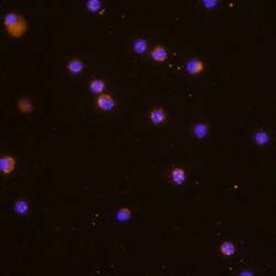
- IL-32 alpha in Human PBMCs. IL-32 alpha was detected in immersion fixed human peripheral blood mononuclear cells (PBMCs) using Human IL-32 alpha Monoclonal Antibody (Catalog # MAB30401) at 10 µg/mL for 3 hours at room temperature. Cells were stained using the NorthernLights™ 557-conjugated Anti-Rat IgG Secondary Antibody (yellow; Catalog # NL013) and counterstained with DAPI (blue). Specific staining was localized to cytoplasm. View our protocol for Fluorescent ICC Staining of Non-adherent Cells.
