 Explore
 Validate
 Learn
Learn Western blot
 Immunocytochemistry
Antibody data
- Antibody Data
- Antigen structure
- References [3]
- Comments [0]
- Validations
- Immunocytochemistry [1]
- Immunohistochemistry [1]
Submit
Validation data
Reference
Comment
Report error
- Product number
- HPA001911 - Provider product page

- Provider
- Atlas Antibodies
- Proper citation
- Atlas Antibodies Cat#HPA001911, RRID:AB_1848427
- Product name
- Anti-FARSA
- Antibody type
- Polyclonal
- Description
- Polyclonal Antibody against Human FARSA, Gene description: phenylalanyl-tRNA synthetase, alpha subunit, Alternative Gene Names: CML33, FARSL, FARSLA, Validated applications: IHC, ICC, WB, Uniprot ID: Q9Y285, Storage: Store at +4°C for short term storage. Long time storage is recommended at -20°C.
- Reactivity
- Human, Mouse, Rat
- Host
- Rabbit
- Conjugate
- Unconjugated
- Isotype
- IgG
- Vial size
- 100 µl
- Concentration
- 0.1 mg/ml
- Storage
- Store at +4°C for short term storage. Long time storage is recommended at -20°C.
- Handling
- The antibody solution should be gently mixed before use.
Submitted references Compound heterozygosity for loss-of-function FARSB
variants in a patient with classic features of recessive aminoacyl-tRNA synthetase-related disease
The proteome of the locus ceruleus in Parkinson's disease: relevance to pathogenesis.
Tissue profiling of the mammalian central nervous system using human antibody-based proteomics.
Antonellis A, Oprescu S, Griffin L, Heider A, Amalfitano A, Innis J
Human Mutation 2018;39(6):834-840
Human Mutation 2018;39(6):834-840
The proteome of the locus ceruleus in Parkinson's disease: relevance to pathogenesis.
van Dijk KD, Berendse HW, Drukarch B, Fratantoni SA, Pham TV, Piersma SR, Huisman E, Brevé JJ, Groenewegen HJ, Jimenez CR, van de Berg WD
Brain pathology (Zurich, Switzerland) 2012 Jul;22(4):485-98
Brain pathology (Zurich, Switzerland) 2012 Jul;22(4):485-98
Tissue profiling of the mammalian central nervous system using human antibody-based proteomics.
Mulder J, Björling E, Jonasson K, Wernérus H, Hober S, Hökfelt T, Uhlén M
Molecular & cellular proteomics : MCP 2009 Jul;8(7):1612-22
Molecular & cellular proteomics : MCP 2009 Jul;8(7):1612-22
No comments: Submit comment
Supportive validation
- Submitted by
- Atlas Antibodies (provider)
- Main image
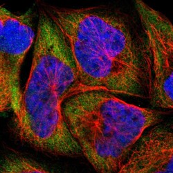
- Experimental details
- Immunofluorescent staining of human cell line A-431 shows localization to cytosol.
- Sample type
- Human

Supportive validation
- Submitted by
- Atlas Antibodies (provider)
- Enhanced method
- Orthogonal validation
- Main image
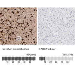
- Experimental details
- Immunohistochemistry analysis in human cerebral cortex and liver tissues using HPA001911 antibody. Corresponding FARSA RNA-seq data are presented for the same tissues.
- Sample type
- Human
- Protocol
- Protocol
