 Explore
 Validate
 Learn
Learn Western blot
 ELISA
ELISAAntibody data
- Antibody Data
- Antigen structure
- References [0]
- Comments [0]
- Validations
- ELISA [1]
- Immunocytochemistry [2]
- Flow cytometry [2]
- Chromatin Immunoprecipitation [2]
- Other assay [1]
Submit
Validation data
Reference
Comment
Report error
- Product number
- 720148 - Provider product page

- Provider
- Invitrogen Antibodies
- Product name
- H2AK119ub Polyclonal Antibody
- Antibody type
- Polyclonal
- Antigen
- Synthetic peptide
- Description
- This antibody is predicted to react with Monkey, Pig and Cat.
- Reactivity
- Human
- Host
- Rabbit
- Isotype
- IgG
- Vial size
- 100 μg
- Concentration
- 0.5 mg/mL
- Storage
- Store at 4°C short term. For long term storage, store at -20°C, avoiding freeze/thaw cycles.
No comments: Submit comment
Supportive validation
- Submitted by
- Invitrogen Antibodies (provider)
- Main image
- Experimental details
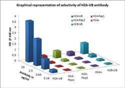
- Cross-reactivity ELISA for Anti-Histone H2A-Ub Rabbit Polyclonal Antibody (Product # 720148) was performed against H2A-Ub, H2A-Pep1, H2A-Pep2, H2Ax and H2B-Ub by coating respective antigens at 20 ng/mL. Antibodies at 10, 2.5, 0.625 and 0.156 µg/mL were added and detected using Goat anti-Rabbit IgG (H+L) Secondary Antibody, HRP conjugate (Product # G-21234, 1:5000 dilution). The plate was developed using Stabilized Chromogen, TMB (Product # SB02).
Supportive validation
- Submitted by
- Invitrogen Antibodies (provider)
- Main image

- Experimental details
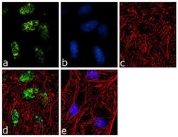
- Immunofluorescence was performed on fixed and permeabilized HeLa cells for detection of H2A-Ub using H2A-Ub Rabbit Polyclonal Antibody (Product # 720148, 2 µg/mL) and labeled with Goat anti-Rabbit IgG (H+L) Superclonal™ Secondary Antibody, Alexa Fluor® 488 conjugate (Product # A27034, 1:2000). Panel a) shows representative cells that were stained for detection and localization of H2A-Ub protein (green), Panel b) is stained for nuclei (blue) using SlowFade® Gold Antifade Mountant with DAPI (Product # S36938). Panel c) represents cytoskeletal F-actin staining using Alexa Fluor® 555 Rhodamine Phalloidin (Product # R415, 1:300). Panel d) is a composite image of Panels a, b and c clearly demonstrating nuclear localization of H2A-Ub. Panel e) represents control cells with no primary antibody to assess background.

- Submitted by
- Invitrogen Antibodies (provider)
- Main image
- Experimental details
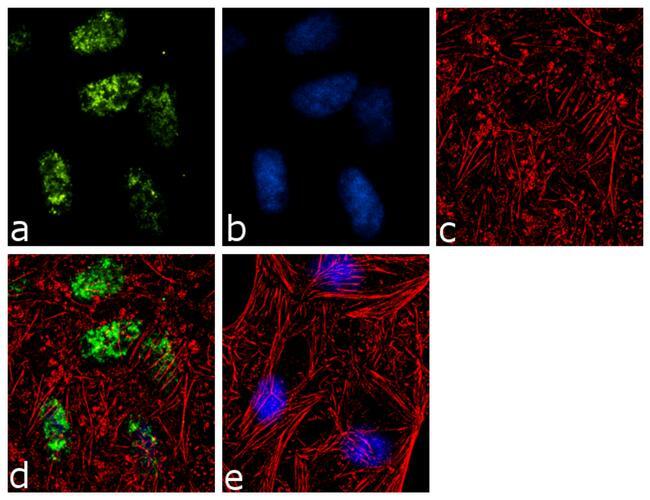
- Immunofluorescence was performed on fixed and permeabilized HeLa cells for detection of H2A-Ub using H2A-Ub Rabbit Polyclonal Antibody (Product # 720148, 2 µg/mL) and labeled with Goat anti-Rabbit IgG (Heavy Chain) Superclonal™ Secondary Antibody, Alexa Fluor® 488 conjugate (Product # A27034, 1:2000). Panel a) shows representative cells that were stained for detection and localization of H2A-Ub protein (green), Panel b) is stained for nuclei (blue) using SlowFade® Gold Antifade Mountant with DAPI (Product # S36938). Panel c) represents cytoskeletal F-actin staining using Alexa Fluor® 555 Rhodamine Phalloidin (Product # R415, 1:300). Panel d) is a composite image of Panels a, b and c clearly demonstrating nuclear localization of H2A-Ub. Panel e) represents control cells with no primary antibody to assess background.
Supportive validation
- Submitted by
- Invitrogen Antibodies (provider)
- Main image

- Experimental details
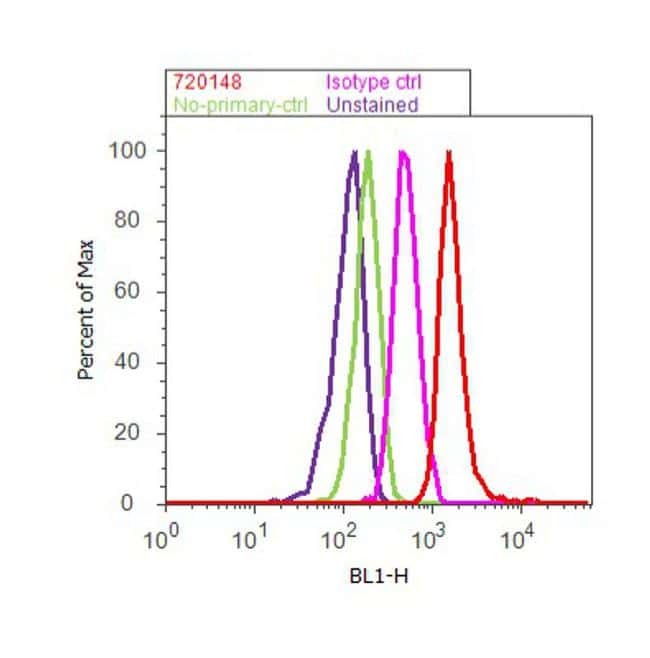
- Flow Cytometry analysis of H2A-Ub was performed on HeLa cells labeled with Anti-H2A-Ub Rabbit Polyclonal Antibody (Product # 720148, 2-4 µg/ 1M cells) or with Rabbit isotype control and detected with Goat anti-Rabbit IgG (H+L) Superclonal™ Secondary Antibody, (Alexa Fluor® 488 conjugate, Product # A27034, 0.4 µg/mL, 1:2500) as represented by the red and pink histograms respectively. The purple histogram represents unstained control cells and the green histogram represents no-primary-Antibody control. A representative of 10,000 cells were acquired and analyzed for each sample using an Attune® Acoustic Focusing Cytometer (Product # 4468770).
- Submitted by
- Invitrogen Antibodies (provider)
- Main image
- Experimental details
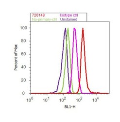
- Flow Cytometry analysis of H2A-Ub was performed on HeLa cells labeled with Anti-H2A-Ub Rabbit Polyclonal Antibody (Product # 720148, 2-4 µg/ 1M cells) or with Rabbit isotype control and detected with Goat anti-Rabbit IgG (Heavy Chain) Superclonal™ Secondary Antibody, (Alexa Fluor® 488 conjugate, Product # A27034, 0.4 µg/mL, 1:2500) as represented by the red and pink histograms respectively. The purple histogram represents unstained control cells and the green histogram represents no-primary-Antibody control. A representative of 10,000 cells were acquired and analyzed for each sample using an Attune® Acoustic Focusing Cytometer (Product # 4468770).

Supportive validation
- Submitted by
- Invitrogen Antibodies (provider)
- Main image
- Experimental details
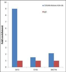
- Enrichment of endogenous Histone H2A-Ub protein using Anti- Histone H2A-Ub Rabbit Polyclonal Antibody: Chromatin Immunoprecipitation (ChIP) was performed using Anti- Histone H2A-Ub Rabbit Polyclonal Antibody (Product # 720148, 3 µg) on sheared chromatin from 2 million HeLa cells using the "MAGnify ChIP system" kit (Product # 49-2024). Normal Rabbit IgG (1 µg) was used as a negative IP control. The purified DNA was analyzed by 7500 Fast qPCR system (Product # 4351106) with optimized PCR primer pairs for the region of inactive SAT2 satellite repeat, used as positive control target, and promoters of the active cFOS, B-Actin region used as negative control target gene. Data is presented as fold enrichment of the antibody signal versus the negative control IgG using the comparative CT method.

- Submitted by
- Invitrogen Antibodies (provider)
- Main image
- Experimental details
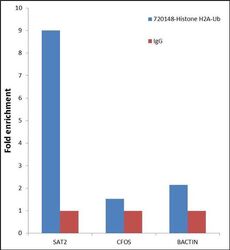
- Enrichment of endogenous Histone H2A-Ub protein using Anti- Histone H2A-Ub Rabbit Polyclonal Antibody: Chromatin Immunoprecipitation (ChIP) was performed using Anti- Histone H2A-Ub Rabbit Polyclonal Antibody (Product # 720148, 3 µg) on sheared chromatin from 2 million HeLa cells using the "MAGnify ChIP system" kit (Product # 49-2024). Normal Rabbit IgG (1 µg) was used as a negative IP control. The purified DNA was analyzed by 7500 Fast qPCR system (Product # 4351106) with optimized PCR primer pairs for the region of inactive SAT2 satellite repeat, used as positive control target, and promoters of the active cFOS, B-Actin region used as negative control target gene. Data is presented as fold enrichment of the antibody signal versus the negative control IgG using the comparative CT method.

Supportive validation
- Submitted by
- Invitrogen Antibodies (provider)
- Main image

- Experimental details
- Cross-reactivity ELISA for Anti-Histone H2A-Ub Rabbit Polyclonal Antibody (Product # 720148) was performed against H2A-Ub, H2A-Pep1, H2A-Pep2, H2Ax and H2B-Ub by coating respective antigens at 20 ng/mL. Antibodies at 10, 2.5, 0.625 and 0.156 µg/mL were added and detected using Goat anti-Rabbit IgG (H+L) Secondary Antibody, HRP conjugate (Product # G-21234, 1:5000 dilution). The plate was developed using Stabilized Chromogen, TMB (Product # SB02).
