Explore
 Validate
 Learn
LearnPA1-16836
antibody from Invitrogen Antibodies
Targeting: BIRC5
API4, EPR-1, survivin
 Western blot
 Immunocytochemistry
 Immunoprecipitation Immunohistochemistry Flow cytometry Chromatin Immunoprecipitation Other assay
Antibody data
- Antibody Data
- Antigen structure
- References [8]
- Comments [0]
- Validations
- Immunocytochemistry [3]
- Immunohistochemistry [2]
- Flow cytometry [1]
- Other assay [9]
Submit
Validation data
Reference
Comment
Report error
- Product number
- PA1-16836 - Provider product page

- Provider
- Invitrogen Antibodies
- Product name
- Survivin Polyclonal Antibody
- Antibody type
- Polyclonal
- Antigen
- Recombinant full-length protein
- Description
- For IHC, prior antigen retrieval (pressure cooking) is recommended for cytoplasmic and nuclear detection of Survivin. Suggested positive control: Hela whole cell extract, antigen standard for BIRC5 (transient overexpression lysate).
- Reactivity
- Human, Mouse, Rat, Canine, Feline, Guinea Pig, Hamster
- Host
- Rabbit
- Isotype
- IgG
- Vial size
- 100 μL
- Concentration
- 1 mg/mL
- Storage
- -20°C or -80°C if preferred
Submitted references Suppression of phosphodiesterase IV enzyme by roflumilast ameliorates cognitive dysfunction in aged rats after sevoflurane anaesthesia via PKA-CREB and MEK/ERK pathways.
In vivo analysis of onset and progression of retinal degeneration in the Nr2e3(rd7/rd7) mouse model of enhanced S-cone sensitivity syndrome.
Pulsed focused ultrasound enhances the therapeutic effect of mesenchymal stromal cell-derived extracellular vesicles in acute kidney injury.
BIRC5/Survivin is a novel ATG12-ATG5 conjugate interactor and an autophagy-induced DNA damage suppressor in human cancer and mouse embryonic fibroblast cells.
Cancer testis antigen expression across T-cell lymphoma subtypes.
The expression of survivin in irreversible pulmonary arterial hypertension rats and its value in evaluating the reversibility of pulmonary arterial hypertension secondary to congenital heart disease.
Hepatitis B virus X protein accelerates hepatocarcinogenesis with partner survivin through modulating miR-520b and HBXIP.
NR4A1 enhances neural survival following oxygen and glucose deprivation: an in vitro study.
Liu P, Wang J, Peng S, Zhang D, Zhuang L, Liu C, Zhang Y, Shi X
The European journal of neuroscience 2022 Aug;56(4):4317-4332
The European journal of neuroscience 2022 Aug;56(4):4317-4332
In vivo analysis of onset and progression of retinal degeneration in the Nr2e3(rd7/rd7) mouse model of enhanced S-cone sensitivity syndrome.
Venturini G, Kokona D, Steiner BL, Bulla EG, Jovanovic J, Zinkernagel MS, Escher P
Scientific reports 2021 Sep 24;11(1):19032
Scientific reports 2021 Sep 24;11(1):19032
Pulsed focused ultrasound enhances the therapeutic effect of mesenchymal stromal cell-derived extracellular vesicles in acute kidney injury.
Ullah M, Liu DD, Rai S, Razavi M, Concepcion W, Thakor AS
Stem cell research & therapy 2020 Sep 14;11(1):398
Stem cell research & therapy 2020 Sep 14;11(1):398
BIRC5/Survivin is a novel ATG12-ATG5 conjugate interactor and an autophagy-induced DNA damage suppressor in human cancer and mouse embryonic fibroblast cells.
Lin TY, Chan HH, Chen SH, Sarvagalla S, Chen PS, Coumar MS, Cheng SM, Chang YC, Lin CH, Leung E, Cheung CHA
Autophagy 2020 Jul;16(7):1296-1313
Autophagy 2020 Jul;16(7):1296-1313
Cancer testis antigen expression across T-cell lymphoma subtypes.
Ma H, Soderquist CR, Marchi E, Scotto L, Bhagat G, O'Connor OA
Hematological oncology 2020 Dec;38(5):827-830
Hematological oncology 2020 Dec;38(5):827-830
The expression of survivin in irreversible pulmonary arterial hypertension rats and its value in evaluating the reversibility of pulmonary arterial hypertension secondary to congenital heart disease.
Li G, Zhang H, Zhao L, Zhang Y, Yan D, Liu Y, Su J, Fan X
Pulmonary circulation 2019 Jul-Sep;9(3):2045894019859480
Pulmonary circulation 2019 Jul-Sep;9(3):2045894019859480
Hepatitis B virus X protein accelerates hepatocarcinogenesis with partner survivin through modulating miR-520b and HBXIP.
Zhang W, Lu Z, Kong G, Gao Y, Wang T, Wang Q, Cai N, Wang H, Liu F, Ye L, Zhang X
Molecular cancer 2014 May 28;13:128
Molecular cancer 2014 May 28;13:128
NR4A1 enhances neural survival following oxygen and glucose deprivation: an in vitro study.
Xiao G, Sun T, Songming C, Cao Y
Journal of the neurological sciences 2013 Jul 15;330(1-2):78-84
Journal of the neurological sciences 2013 Jul 15;330(1-2):78-84
No comments: Submit comment
Supportive validation
- Submitted by
- Invitrogen Antibodies (provider)
- Main image
- Experimental details
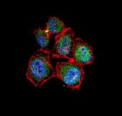
- Immunocytochemistry analysis of Survivin in HeLa cells. Samples were incubated in Survivin polyclonal antibody (Product # PA1-16836) using a dilution of 1:10 followed by Alexa Fluor 488-conjugated Goat to rabbit IgG secondary antibody (green). Actin filaments were labeled with Alexa Fluor 568 phalloidin (red). DAPI was used to stain the cell nuclei (blue).

- Submitted by
- Invitrogen Antibodies (provider)
- Main image

- Experimental details
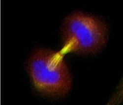
- Immunocytochemistry analysis of Survivin in Telophase with accumulation of survivin in the midbodies of two daughter cells. Samples were incubated in Survivin polyclonal antibody (Product # PA1-16836). Analysis using the HRP conjugate.

- Submitted by
- Invitrogen Antibodies (provider)
- Main image
- Experimental details
- Immunocytochemistry analysis of Survivin in Telophase with accumulation of survivin in the midbodies of two daughter cells. Samples were incubated in Survivin polyclonal antibody (Product # PA1-16836). Analysis using the HRP conjugate.

Supportive validation
- Submitted by
- Invitrogen Antibodies (provider)
- Main image
- Experimental details
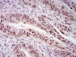
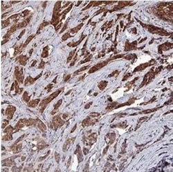
- Immunohistochemical analysis of Survivin in human rectal cancer. Samples were incubated in Survivin polyclonal antibody (Product # PA1-16836) followed by DAB with hematoxylin counterstain.

- Submitted by
- Invitrogen Antibodies (provider)
- Main image
- Experimental details
- Immunohistochemical analysis of Survivin in lysates of human neuroblastoma cell line. Samples were incubated in Survivin polyclonal antibody (Product # PA1-16836) using a dilution of 1:200 followed by a goat anti-rabbit IgG at a dilution of 1:2000.

Supportive validation
- Submitted by
- Invitrogen Antibodies (provider)
- Main image
- Experimental details
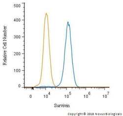
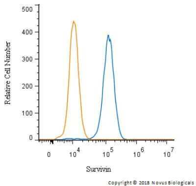
- Flow cytometry of Survivin in HeLa cells with and a matched isotype control. Samples were incubated in Survivin polyclonal antibody (Product # PA1-16836) using a dilution of 2.5 µg/mL for 30 minutes at room temperature followed by a Rabbit IgG (H+L) Cross-Adsorbed Secondary Antibody, Dylight™ 550. Cells were fixed with 4% PFA and then permeabilized with 0.1% saponin.
Supportive validation
- Submitted by
- Invitrogen Antibodies (provider)
- Main image

- Experimental details
- NULL
- Submitted by
- Invitrogen Antibodies (provider)
- Main image
- Experimental details
- NULL

- Submitted by
- Invitrogen Antibodies (provider)
- Main image
- Experimental details
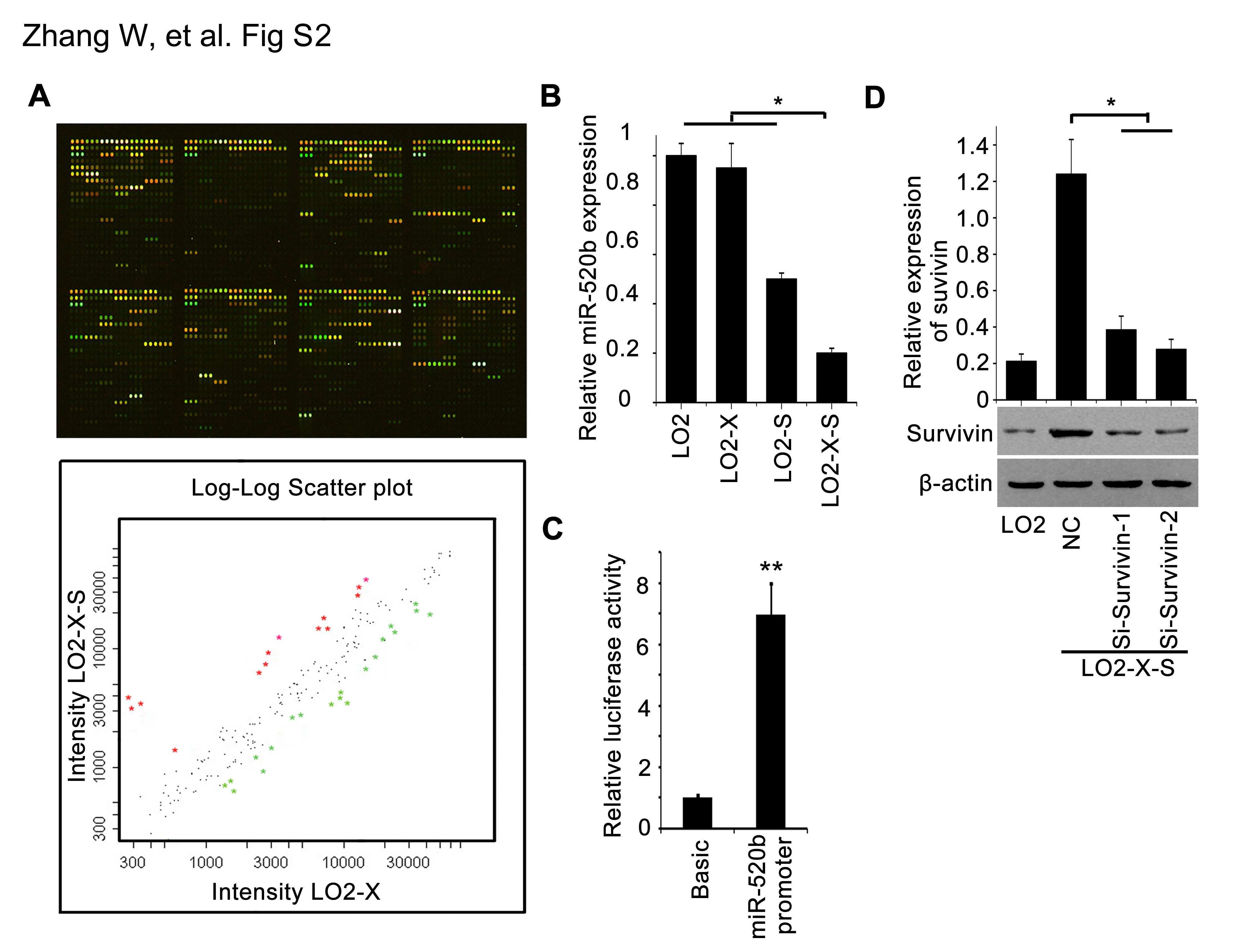
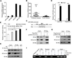
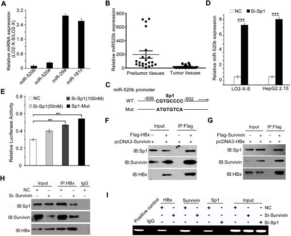
- Figure 2 HBx down-regulates miR-520b through binding to Sp1 with partner survivin. (A) The expression levels of miRNA-520b, miRNA-520e, miRNA-29a and miRNA-181c were examined by qRT-PCR in LO2-X-S/LO2-X cells. (B) The expression of miR-520b in clinical HCC and peritumor samples was detected by qRT-PCR. (C) A model shows Sp1 binding site-directed mutation in the promoter region of miR-520b. (D) The effect of knockdown of Sp1 on miR-520b in LO2-X-S or HepG2.2.15 cells was examined by qRT-PCR analysis (*** P < 0.001, Student's t test). (E) The effect of Sp1 on miR-520b promoter in LO2-X-S cells was tested using Sp1 siRNA (Si-Sp1) or Sp1 mutant by luciferase reporter gene assays (** P < 0.01, Student's t test). (F-H) The interaction among HBx, survivin and Sp1 in a complex was examined by co-IP. (I) Interaction of the complex, including HBx, survivin and Sp1, with the promoter region of miR-520b was examined by ChIP in LO2-X-S cells.
- Submitted by
- Invitrogen Antibodies (provider)
- Main image
- Experimental details
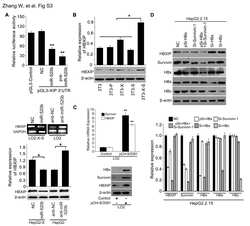
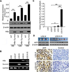
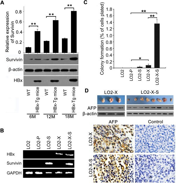
- Figure 1 HBx accelerates hepatocarcinogenesis with partner survivin. (A) The expression of survivin in the liver tissues of p21-HBx Tg mice aged 6, 12 and 18 mouths versus WT littermate mice were determined by western blotting, respectively (** P < 0.01, Student's t test). (B) The integrations of HBx and survivin genes into the genomes of LO2 cells were validated by PCR using genomic DNA as a template. GAPDH was used as a loading control. (C) The effect of HBx and/or survivin on cell proliferation was detected by colony-formation assay (* P < 0.05, ** P < 0.01, Student's t test). (D) Tumor formation in nude mice (n = 8 per group) injected with LO2-X or LO2-X-S cells was assessed in 3 weeks. The expression of AFP was tested in the tumor tissues from mice by western blotting and IHC analysis, respectively.
- Submitted by
- Invitrogen Antibodies (provider)
- Main image

- Experimental details
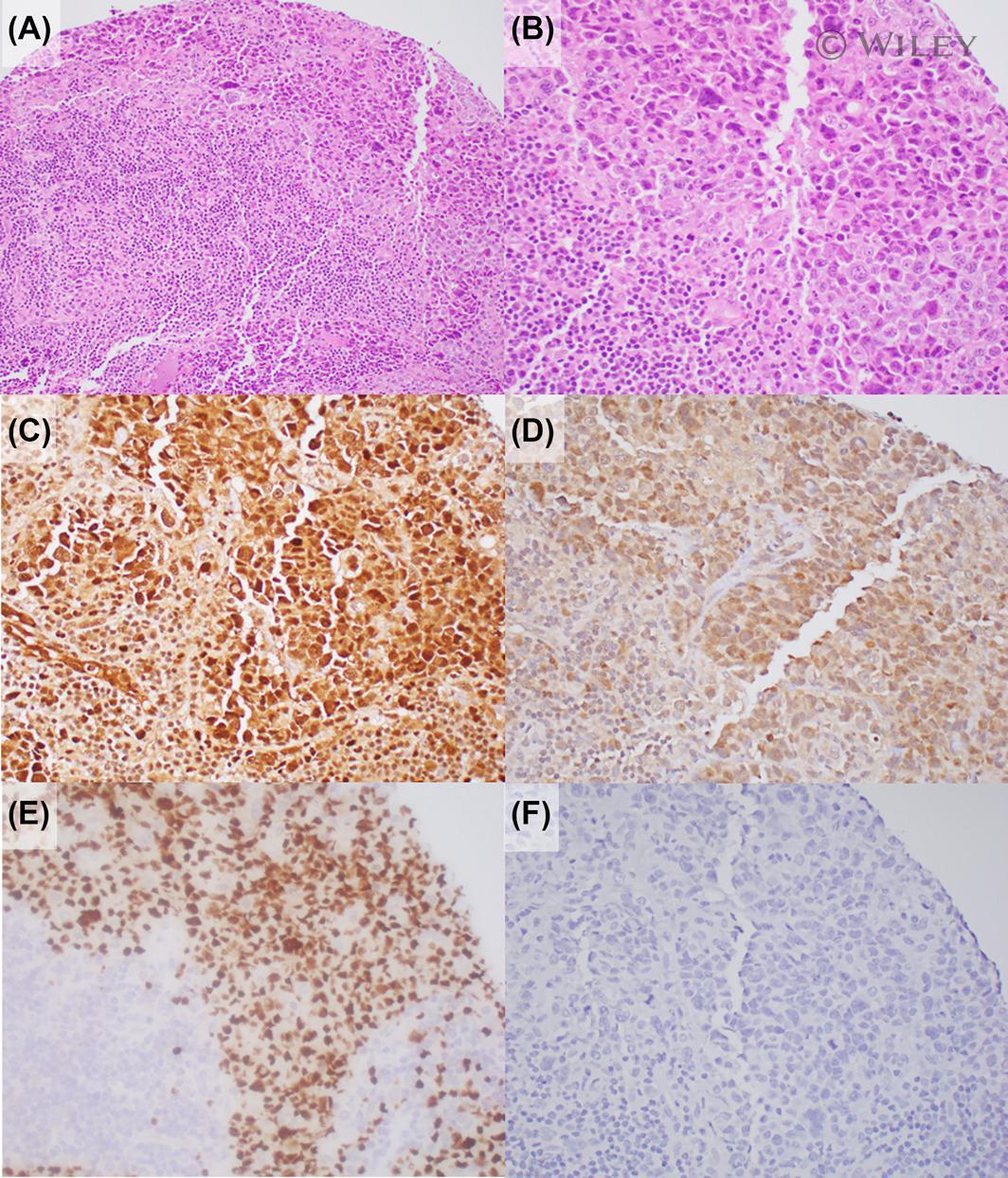
- 1 FIGURE Representative expression of cancer testis antigens by immunohistochemistry in a patient with ALK-negative anaplastic large cell lymphoma. Panels A and B demonstrate hematoxylin and eosin staining at lower (20x) and higher power (40x). Panel C shows survivin staining at 75%-100% expression with strong intensity, localized in both the nucleus and cytoplasm. Panel D shows SCP1 expressed in 50%-75% of cells with weak intensity, localized in both the nucleus and cytoplasm. Panel E shows WT1 positivity, localized in the nucleus, in 50%-75% of cells with strong intensity. Panel F shows negative staining for PLAC1, but this was also representative of PRAME and SSX2
- Submitted by
- Invitrogen Antibodies (provider)
- Main image

- Experimental details
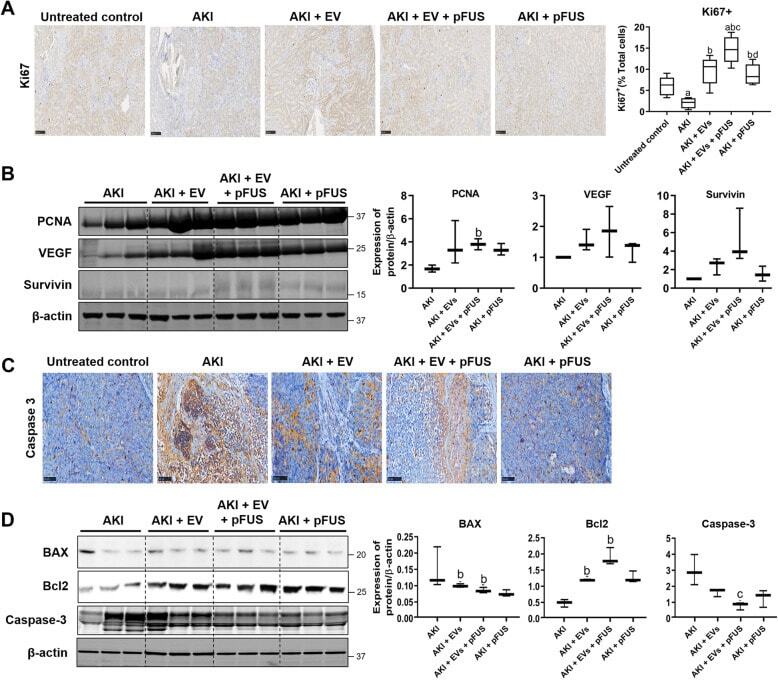
- Fig. 5 Proliferation and apoptosis. a Immunohistochemical staining for proliferation marker Ki67 in kidney tissue (left), alongside quantification of the percentage of Ki67-positive cells (right). b Western blot on kidney tissue measuring proliferation markers PCNA, VEGF, survivin, and beta-actin (left) alongside quantification (right). c Immunohistochemical staining for apoptosis marker Caspase-3 in kidney tissue. d Western blot on kidney tissue measuring apoptosis markers BAX, Bcl-2, and Caspase-3 (left) alongside quantification (right). Each group has n = 3 mice. Significant difference a p < 0.05: relative to untreated control; b p < 0.05: relative to AKI; c p < 0.05: relative to AKI-EV; d p < 0.05: relative to AKI-EV-pFUS
- Submitted by
- Invitrogen Antibodies (provider)
- Main image
- Experimental details
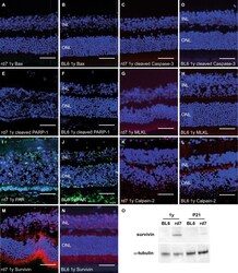
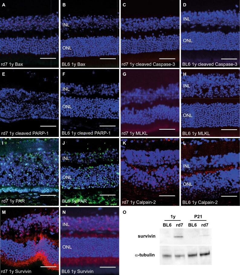
- Figure 6 Cell death mechanisms in C57BL/6J Nr2e3 rd7/rd7 retinas. Immunohistochemical analysis of cell death markers on 5 muM-paraffin sections of 1-year-old (1y) C57BL/6J Nr2e3 rd7/rd7 (rd7) ( A , C , E , G , I , K , M ) and wild-type C57BL/6J (BL6) ( B , D , F , H , J , L , N ) retinas. Sections were probed with antibodies raised against Bax ( A , B ), cleaved Caspase-3 ( C , D ), cleaved PARP-1 ( E , F ), MLKL ( G , H ), PAR ( I , J ), Calpain-2 ( K , L ) and Survivin ( M , N ). Bax, cleaved Caspase-3, MLKL and Survivin were detected with a secondary antibody conjugated to Alexa Fluor 594 (red), cleaved PARP-1 and Calpain-2 with a secondary antibody conjugated to Cy5 (red) and PAR with a secondary antibody conjugated to FITC (green). Nuclei were stained with DAPI (blue), namely the outer nuclear layer (ONL) and the inner nuclear layer (INL). Scale bar: 50 um. ( O ) Qualitative Western blot analysis on six pooled retinas of 21-day (P21) and 1-year-old (1y) C57BL/6J (BL6) and C57BL/6J Nr2e3 rd7/rd7 (rd7) mice. Expression of the 16-kDa survivin and 49-kDa alpha-tubulin proteins were assessed.
- Submitted by
- Invitrogen Antibodies (provider)
- Main image
- Experimental details
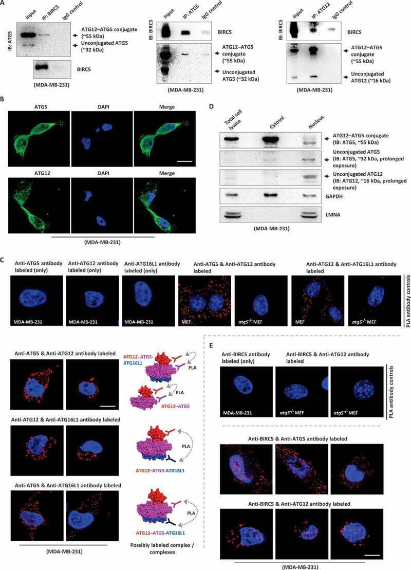
- Figure 5. BIRC5 interacts with ATG12-ATG5 conjugate in cancer cells. (A) Lysates of MDA-MB-231 cells were immunoprecipitated with anti-BIRC5, anti-ATG5, or anti-ATG12 antibodies. Protein-protein interactions between BIRC5, ATG12-ATG5 conjugate, ATG12, and ATG5 were determined by western blotting. (B) Expression of ATG5/ATG5-containing protein complexes and ATG12/ATG12-containing protein complexes was visualized by immunofluorescent microscopy. Nucleus were countered stained blue by DAPI. (C) Endogenous physical interactions between the examined molecules in MDA-MB-231 cells were detected by in situ PLA (indicated by red fluorescent puncta) and visualized by fluorescence microscopy. The Atg12-Atg5 conjugate deficient atg5 -/- MEF cells were used as an antibody-specificity control for the ATG12-ATG5 and ATG12-ATG16L1 PLA assays. Nucleus were counter stained blue by DAPI. (D) Cytoplasmic and nucleic proteins were isolated and extracted from MDA-MB-231 cells. The present of ATG12-ATG5 conjugate, ATG12, and ATG5 in the extracted cytoplasmic and nucleic protein fractions was determined by western blotting. GAPDH/Gapdh and LMNA (lamin A/C) were used as the internal control of the cytoplasmic and nucleic protein fraction, respectively. (E) Endogenous physical interactions between BIRC5, ATG5, and ATG12 in MDA-MB-231 cells were detected by in situ PLA (indicated by red fluorescent puncta) and visualized by fluorescent microscopy. Nucleus were counter stained blue by DAPI. Scale bars:
- Submitted by
- Invitrogen Antibodies (provider)
- Main image

- Experimental details
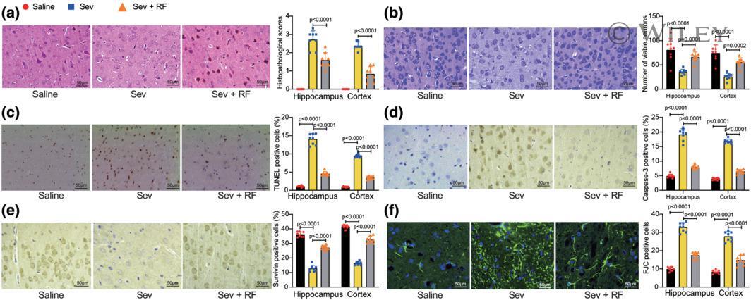
- 2 FIGURE RF prevents neurons from apoptosis in Sev-induced aged rats. (a) Pathological damage in hippocampus and cortex of rats by HE staining ( F 2,42 = 634.9, P < 0.0001); (b) neuronal morphology and viability in hippocampus and cortex by Nissl staining ( F 2,42 = 50.01, P < 0.0001); (c) TUNEL staining of apoptotic cells in hippocampus and cortex of rats ( F 2, 42 = 970.6, P < 0.0001); (d) the number of caspase-3-positive cells in hippocampus and cortex of rats by immunohistochemistry ( F 2,42 = 821.0, P < 0.0001); (e) the number of survivin-positive cells in hippocampus and cortex of rats by immunohistochemistry ( F 2,41 = 757.0, P < 0.0001); (f) the number of degenerative neurons in hippocampus and cortex of rats by FJC staining ( F 2,42 = 503.0, P < 0.0001). Each group contained eight rats. The behavioural experiments were performed by at least two experimenters under double-blind conditions. All experiments were performed as three independent sets in triplicate. The results are presented as the mean +- SD . Two-way ANOVA followed by Tukey's post hoc test was used to perform comparisons among multiple groups.