Explore
 Validate
 Learn
Learn Western blot
 Immunohistochemistry
Antibody data
- Antibody Data
- Antigen structure
- References [2]
- Comments [0]
- Validations
- Immunohistochemistry [1]
Submit
Validation data
Reference
Comment
Report error
- Product number
- AF3385 - Provider product page

- Provider
- Novus Biologicals
- Product name
- Goat Polyclonal Nidogen-2 Antibody
- Antibody type
- Polyclonal
- Description
- Antigen Affinity-purified. Detects human Nidogen-2 in direct ELISAs and Western blots. In direct ELISAs and Western blots, less than 1% cross-reactivity with recombinant human Nidogen-1 is observed.
- Reactivity
- Human
- Host
- Goat
- Conjugate
- Unconjugated
- Isotype
- IgG
- Vial size
- 100 ug
- Concentration
- LYOPH
- Storage
- Use a manual defrost freezer and avoid repeated freeze-thaw cycles. 12 months from date of receipt, -20 to -70 degreesC as supplied. 1 month, 2 to 8 degreesC under sterile conditions after reconstitution. 6 months, -20 to -70 degreesC under sterile conditions after reconstitution.
Submitted references Hematopoietic stem cell cytokines and fibroblast growth factor-2 stimulate human endothelial cell-pericyte tube co-assembly in 3D fibrin matrices under serum-free defined conditions.
Pericyte recruitment during vasculogenic tube assembly stimulates endothelial basement membrane matrix formation.
Smith AO, Bowers SL, Stratman AN, Davis GE
PloS one 2013;8(12):e85147
PloS one 2013;8(12):e85147
Pericyte recruitment during vasculogenic tube assembly stimulates endothelial basement membrane matrix formation.
Stratman AN, Malotte KM, Mahan RD, Davis MJ, Davis GE
Blood 2009 Dec 3;114(24):5091-101
Blood 2009 Dec 3;114(24):5091-101
No comments: Submit comment
Supportive validation
- Submitted by
- Novus Biologicals (provider)
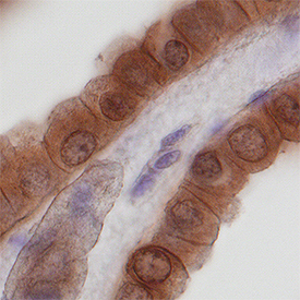
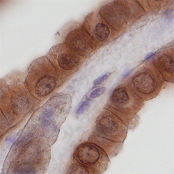
- Main image
- Experimental details
- Nidogen-2 in Human Kidney. Nidogen-2 was detected in immersion fixed paraffin-embedded sections of normal human kidney using Goat Anti-Human Nidogen-2 Antigen Affinity-purified Polyclonal Antibody (Catalog # AF3385) at 10 µg/mL overnight at 4 °C. Tissue was stained using the Anti-Goat HRP-DAB Cell & Tissue Staining Kit (brown; Catalog # CTS008) and counterstained with hematoxylin (blue). Specific staining was localized to epithelial cells in convoluted tubules. View our protocol for Chromogenic IHC Staining of Paraffin-embedded Tissue Sections.