 Explore
 Validate
 Learn
Learn Western blot
 Immunohistochemistry
Antibody data
- Antibody Data
- Antigen structure
- References [0]
- Comments [0]
- Validations
- Western blot [1]
- Immunocytochemistry [1]
- Immunohistochemistry [8]
Submit
Validation data
Reference
Comment
Report error
- Product number
- HPA052460 - Provider product page

- Provider
- Atlas Antibodies
- Proper citation
- Atlas Antibodies Cat#HPA052460, RRID:AB_2681840
- Product name
- Anti-NID2
- Antibody type
- Polyclonal
- Reactivity
- Human
- Host
- Rabbit
- Conjugate
- Unconjugated
- Antigen sequence
HTKEGTSLGEVGGPDLKGQVEPWDERETRSPAPPE
VDRDSLAPSWETPPPYPENGSIQPYPDGGPVPSEM
DVPPAHPEEEIVLRSYPASGHTT- Isotype
- IgG
- Vial size
- 100 µl
- Storage
- Store at +4°C for short term storage. Long time storage is recommended at -20°C.
No comments: Submit comment
Supportive validation
- Submitted by
- Atlas Antibodies (provider)
- Enhanced method
- Independent antibody validation
- Main image
- Experimental details
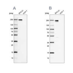
- Western blot analysis using Anti-NID2 antibody HPA052460 (A) shows similar pattern to independent antibody HPA058772 (B).

Supportive validation
- Submitted by
- Atlas Antibodies (provider)
- Main image

- Experimental details
- Immunofluorescent staining of human cell line BJ shows localization to plasma membrane.
- Sample type
- HUMAN

Enhanced validation
Supportive validation
- Submitted by
- Atlas Antibodies (provider)
- Enhanced method
- Orthogonal validation
- Main image
- Experimental details
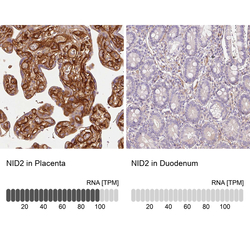
- Immunohistochemistry analysis in human placenta and duodenum tissues using HPA052460 antibody. Corresponding NID2 RNA-seq data are presented for the same tissues.
- Sample type
- HUMAN

Supportive validation
- Submitted by
- Atlas Antibodies (provider)
- Main image
- Experimental details
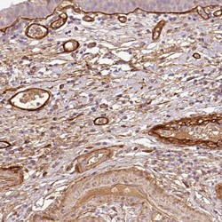
- Immunohistochemical staining of human urinary bladder shows distinct positivity in connective tissue as well as basement membrane.

- Submitted by
- Atlas Antibodies (provider)
- Main image

- Experimental details
- Immunohistochemical staining of human placenta shows high expression.
- Sample type
- HUMAN

- Submitted by
- Atlas Antibodies (provider)
- Main image
- Experimental details
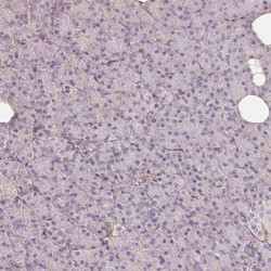
- Immunohistochemical staining of human pancreas shows low expression as expected.
- Sample type
- HUMAN

- Submitted by
- Atlas Antibodies (provider)
- Main image
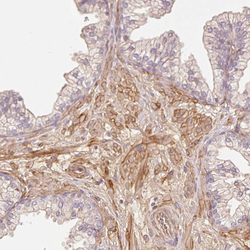
- Experimental details
- Immunohistochemical staining of human prostate shows moderate membranous positivity in smooth muscle cells.
- Sample type
- HUMAN

- Submitted by
- Atlas Antibodies (provider)
- Main image
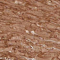
- Experimental details
- Immunohistochemical staining of human heart muscle shows strong membranous positivity in cardiomyocytes.
- Sample type
- HUMAN

- Submitted by
- Atlas Antibodies (provider)
- Main image
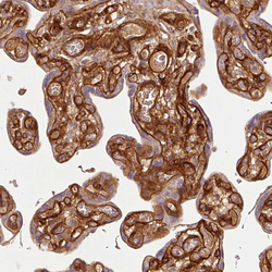
- Experimental details
- Immunohistochemical staining of human placenta shows strong membranous positivity in trophoblastic cells.
- Sample type
- HUMAN

- Submitted by
- Atlas Antibodies (provider)
- Main image
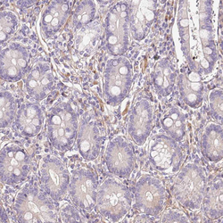
- Experimental details
- Immunohistochemical staining of human duodenum shows no positivity in glandular cells as expected.
- Sample type
- HUMAN
