Explore
 Validate
 Learn
Learn Western blot
Antibody data
- Antibody Data
- Antigen structure
- References [2]
- Comments [0]
- Validations
- Western blot [3]
Submit
Validation data
Reference
Comment
Report error
- Product number
- AF4177 - Provider product page

- Provider
- R&D Systems
- Product name
- Human/Mouse/Rat PKA RI beta Antibody
- Antibody type
- Polyclonal
- Description
- Immunogen affinity purified. Detects human, mouse, and rat PKA RI beta in Western blots. In Western blots, less than 1% cross-reactivity with recombinant human (rh) PKA RI alpha or rhPKA RII beta is observed; cross-reactivity with PKA RII alpha is unknown.
- Reactivity
- Human, Mouse, Rat
- Host
- Sheep
- Conjugate
- Unconjugated
- Antigen sequence
P31321- Isotype
- IgG
- Vial size
- 100 ug
- Concentration
- LYOPH
- Storage
- Use a manual defrost freezer and avoid repeated freeze-thaw cycles. 12 months from date of receipt, -20 to -70 °C as supplied. 1 month, 2 to 8 °C under sterile conditions after reconstitution. 6 months, -20 to -70 °C under sterile conditions after reconstitution.
Submitted references A Bayesian Framework for Generalized Linear Mixed Modeling Identifies New Candidate Loci for Late-Onset Alzheimer's Disease.
microRNA-208a in an early stage myocardial infarction rat model and the effect on cAMP-PKA signaling pathway.
Wang X, Philip VM, Ananda G, White CC, Malhotra A, Michalski PJ, Karuturi KRM, Chintalapudi SR, Acklin C, Sasner M, Bennett DA, De Jager PL, Howell GR, Carter GW
Genetics 2018 May;209(1):51-64
Genetics 2018 May;209(1):51-64
microRNA-208a in an early stage myocardial infarction rat model and the effect on cAMP-PKA signaling pathway.
Feng G, Yan Z, Li C, Hou Y
Molecular medicine reports 2016 Aug;14(2):1631-5
Molecular medicine reports 2016 Aug;14(2):1631-5
No comments: Submit comment
Supportive validation
- Submitted by
- R&D Systems (provider)
- Main image
- Experimental details
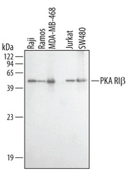
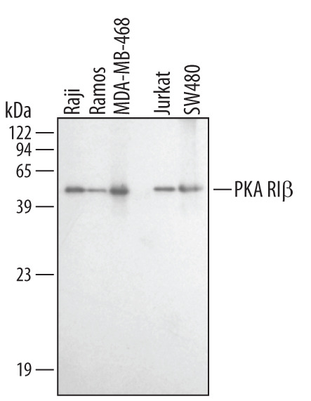
- Detection of Human PKA RI beta by Western Blot. Western blot shows lysates of MDA-MB-468 human breast cancer cell line, Jurkat human acute T cell leukemia cell line, SW480 human colorectal adenocarcinoma cell line, Raji human Burkitt's lymphoma cell line, and Ramos human Burkitt's lymphoma cell line. PVDF membrane was probed with 1 µg/mL Sheep Anti-Human/Mouse/Rat PKA RI beta Antigen Affinity-purified Polyclonal Antibody (Catalog # AF4177) followed by HRP-conjugated Anti-Sheep IgG Secondary Antibody (Catalog # HAF016). A specific band for PKA RI beta was detected at approximately 48 kDa (as indicated). This experiment was conducted under reducing conditions and using Immunoblot Buffer Group 1.
- Submitted by
- R&D Systems (provider)
- Main image
- Experimental details
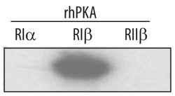
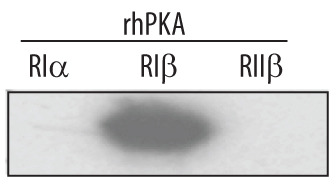
- Detection of Human PKA RI beta by Western Blot. Western blot shows recombinant human PKA RI alpha , PKA RI beta , and PKA RII beta (5 ng/lane). PVDF membrane was probed with 1 µg/mL Sheep Anti-Human/ Mouse/Rat PKA RI beta Antigen Affinity-purified Polyclonal Antibody (Catalog # AF4177) followed by HRP-conjugated Anti-Sheep IgG Secondary Antibody (Catalog # HAF016). A specific band for PKA RI beta was detected at approximately 48 kDa (as indicated). This experiment was conducted under reducing conditions and using Immunoblot Buffer Group 1.
- Submitted by
- R&D Systems (provider)
- Main image
- Experimental details
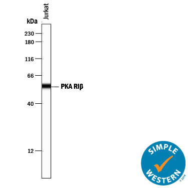
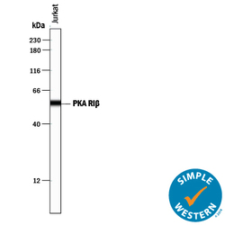
- Detection of Human PKA RI beta by Simple WesternTM. Simple Western lane view shows lysates of Jurkat human acute T cell leukemia cell line, loaded at 0.2 mg/mL. A specific band was detected for PKA RI beta at approximately 56 kDa (as indicated) using 10 µg/mL of Sheep Anti-Human/Mouse/Rat PKA RI beta Antigen Affinity-purified Polyclonal Antibody (Catalog # AF4177) followed by 1:50 dilution of HRP-conjugated Anti-Sheep IgG Secondary Antibody (Catalog # HAF016). This experiment was conducted under reducing conditions and using the 12-230 kDa separation system.