 Explore
 Validate
 Learn
Learn Western blot
 Immunoprecipitation
Antibody data
- Antibody Data
- Antigen structure
- References [2]
- Comments [0]
- Validations
- Western blot [3]
- Immunocytochemistry [1]
- Other assay [2]
Submit
Validation data
Reference
Comment
Report error
- Product number
- PA5-17325 - Provider product page

- Provider
- Invitrogen Antibodies
- Product name
- Prohibitin Polyclonal Antibody
- Antibody type
- Polyclonal
- Antigen
- Synthetic peptide
- Description
- It is not recommended to aliquot this antibody.
- Reactivity
- Human, Mouse, Rat
- Host
- Rabbit
- Isotype
- IgG
- Vial size
- 100 µL
- Storage
- -20°C
Submitted references Nuclear partitioning of Prohibitin 1 inhibits Wnt/β-catenin-dependent intestinal tumorigenesis.
Neuronal Pentraxin 1 Promotes Hypoxic-Ischemic Neuronal Injury by Impairing Mitochondrial Biogenesis via Interactions With Active Bax[6A7] and Mitochondrial Hexokinase II.
Alula KM, Delgado-Deida Y, Jackson DN, Venuprasad K, Theiss AL
Oncogene 2021 Jan;40(2):369-383
Oncogene 2021 Jan;40(2):369-383
Neuronal Pentraxin 1 Promotes Hypoxic-Ischemic Neuronal Injury by Impairing Mitochondrial Biogenesis via Interactions With Active Bax[6A7] and Mitochondrial Hexokinase II.
Al Rahim M, Thatipamula S, Pasinetti GM, Hossain MA
ASN neuro 2021 Jan-Dec;13:17590914211012888
ASN neuro 2021 Jan-Dec;13:17590914211012888
No comments: Submit comment
Supportive validation
- Submitted by
- Invitrogen Antibodies (provider)
- Main image

- Experimental details
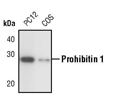
- Western blot analysis of PHB1 in extracts from PC12 and COS cells using PHB1 polyclonal antibody (Product # PA5-17325).
- Submitted by
- Invitrogen Antibodies (provider)
- Main image

- Experimental details
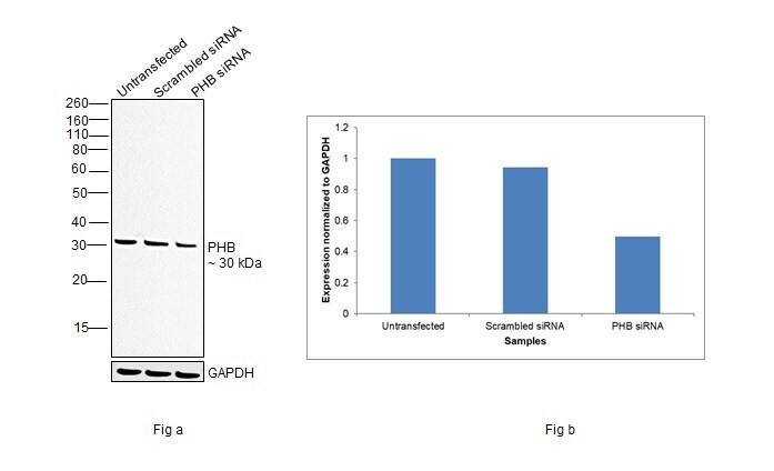
- Knockdown of Prohibitin was achieved by transfecting A-431 with Prohibitin specific siRNAs (Silencer® select Product # s10425, s10424). Western blot analysis (Fig. a) was performed using Whole cell extracts from the Prohibitin knockdown cells (lane 3), non-targeting scrambled siRNA transfected cells (lane 2) and untransfected cells (lane 1). The blot was probed with Prohibitin Polyclonal Antibody (Product # PA5-17325, 1:1000 dilution) and Goat anti-Rabbit IgG (H+L) Superclonal™ Recombinant Secondary Antibody, HRP (Product # A27036, 1:4000 dilution). Densitometric analysis of this western blot is shown in histogram (Fig. b). Decrease in signal upon siRNA mediated knock down confirms that antibody is specific to Prohibitin.
- Submitted by
- Invitrogen Antibodies (provider)
- Main image
- Experimental details
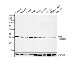
- Western blot was performed using Anti-Prohibitin Polyclonal Antibody(Product # PA5-17325) and a 30 kDa band corresponding to Prohibitin was observed across cell lines and tissues tested. Whole cell extracts (30 µg lysate) of Caco-2 (Lane 1), Hep G2 (Lane 2), T-47D (Lane 3), A-431 (Lane 4), HeLa (Lane 5), NIH/3T3 (Lane 6), PC-12 (Lane 7) and tissue extracts (30 µg) of Mouse Heart (Lane 8), Rat Brain (Lane 9) were electrophoresed using NuPAGE™ 12% Bis-Tris Protein Gel (Product # NP0341BOX). Resolved proteins were then transferred onto a Nitrocellulose membrane (Product # IB23001) by iBlot® 2 Dry Blotting System (Product # IB21001). The blot was probed with the primary antibody (1:1000 dilution) and detected by chemiluminescence with Goat anti-Rabbit IgG (H+L) Superclonal™ Recombinant Secondary Antibody, HRP (Product # A27036,1:4000 dilution) using the iBright FL 1000 (Product # A32752). Chemiluminescent detection was performed using Novex® ECL Chemiluminescent Substrate Reagent Kit (Product # WP20005).

Supportive validation
- Submitted by
- Invitrogen Antibodies (provider)
- Main image

- Experimental details
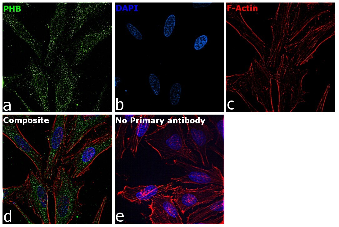
- Immunofluorescence analysis of Prohibitin was performed using 70% confluent log phase HeLa cells. The cells were fixed with 4% paraformaldehyde for 10 minutes, permeabilized with 0.1% Triton™ X-100 for 45 minutes, and blocked with 2% BSA for 45 minutes at room temperature. The cells were labeled with Prohibitin Polyclonal Antibody (Product # PA5-17325) at 1:100 dilution in 0.1% BSA, incubated at 4 degree celsius overnight and then labeled with Goat anti-Rabbit IgG (H+L) Highly Cross-Adsorbed Secondary Antibody, Alexa Fluor Plus 488 (Product # A32731), (1:2000 dilution), for 45 minutes at room temperature (Panel a: Green). Nuclei (Panel b:Blue) were stained with SlowFade® Gold Antifade Mountant with DAPI (Product # S36938). F-actin (Panel c: Red) was stained with Rhodamine Phalloidin (Product # R415, 1:300 dilution). Panel d represents the merged image showing cytoplasm and nucleus localization. Panel e represents control cells with no primary antibody to assess background. The images were captured at 60X magnification.
Supportive validation
- Submitted by
- Invitrogen Antibodies (provider)
- Main image

- Experimental details
- Figure 1. PHB1 expression inversely correlates with beta-catenin expression in adenomas. Western blots of nuclear and cytosolic protein fractions of isolated IECs from jejunum (A) or mucosa of colon (B) of wild-type (WT) and Apc Min/+ mice or isolated adenomas from Apc Min/+ mice. *denotes non-specific band in (A), likely family member PHB2. Mean +- SEM of PHB1 western blot densitometry in jejunum (C) or colon (D). Mean +- SEM of beta-catenin western blot densitometry in jejunum (E) or colon (F). (G) Spearman rank correlation of western densitometry in nuclear fraction of adenomas. n = 3 mice per group. * P < 0.05, ** P < 0.01 vs. WT IECs by one-way ANOVA followed by Bonferroni's test.

- Submitted by
- Invitrogen Antibodies (provider)
- Main image
- Experimental details
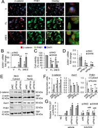
- Figure 8. Relative expression of PHB1 regulates beta-catenin activation and cell viability in RKO and SW48 CRC cell lines. (A-C) RKO or SW48 cells were transfected with pCDNA4 (vector; V), full-length pCDNA4-PHB1 (FL), or pCDNA4-PHB1 DeltaNES (DeltaNES) for 72 hr. (A) Immunofluorescent staining of RKO cells. Scale bars: 20 mum; boxed pullouts: 20 mum. (B) Mean +- SEM of relative AXIN1 mRNA expression. (C) Cell viability as measured by LDH release. (D-G) RKO or SW48 cells were transfected with 2 unique siRNAs against PHB1 or siNegative Control (siNC) for 48 hr. (D) Mean +- SEM of relative AXIN1 mRNA expression. (E) Representative western blot of beta-catenin, AXIN1, or PHB1 (to demonstrate efficiency of siRNA knockdown). (F) Mean +- SEM of densitometric units by western blotting. (G) Relative luciferase expression of transcriptional activation by beta-Catenin after 10 muM XAV939 or vehicle treatment for 16 hr. Negative control (NC) cells were transfected with a non-inducible firefly reporter construct. n = 3 (A, D-G) or n = 6 (B, C) biological replicates per group. * P < 0.05, ** P < 0.01 vs V by unpaired, two-tailed Student's t test (B, C); * P < 0.05 vs siNC vehicle, # P < 0.05, ## P < 0.01 vs siPhb1 vehicle by one-way ANOVA (D, F) or two-way ANOVA followed by Bonferroni's test (G).
