Explore
 Validate
 Learn
Learn Western blot
 Immunoprecipitation
Antibody data
- Antibody Data
- Antigen structure
- References [3]
- Comments [0]
- Validations
- Western blot [4]
- Immunocytochemistry [2]
- Immunohistochemistry [8]
- Other assay [4]
Submit
Validation data
Reference
Comment
Report error
- Product number
- PA5-27329 - Provider product page

- Provider
- Invitrogen Antibodies
- Product name
- Prohibitin Polyclonal Antibody
- Antibody type
- Polyclonal
- Antigen
- Recombinant full-length protein
- Description
- Recommended positive controls: 293T, A431, HeLa, HepG2, NIH-3T3, PC-12. Predicted reactivity: Mouse (99%), Rat (99%), Zebrafish (93%), Xenopus laevis (92%), Dog (100%), Cat (100%), Chicken (95%), Rhesus Monkey (100%), Chimpanzee (100%), Bovine (100%). Store product as a concentrated solution. Centrifuge briefly prior to opening the vial.
- Reactivity
- Human, Mouse, Rat
- Host
- Rabbit
- Isotype
- IgG
- Vial size
- 100 μL
- Concentration
- 0.34 mg/mL
- Storage
- Store at 4°C short term. For long term storage, store at -20°C, avoiding freeze/thaw cycles.
Submitted references The Pseudomonas aeruginosa Type III Secretion System Exoenzyme Effector ExoU Induces Mitochondrial Damage in a Murine Bone Marrow-Derived Macrophage Infection Model.
Cutting Edge: Mitochondrial Assembly of the NLRP3 Inflammasome Complex Is Initiated at Priming.
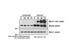
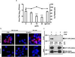
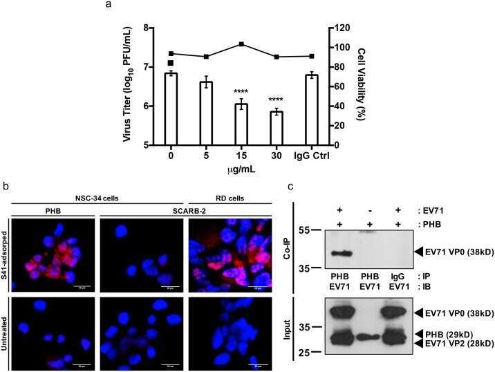
Prohibitin plays a critical role in Enterovirus 71 neuropathogenesis.
Hardy KS, Tuckey AN, Housley NA, Andrews J, Patel M, Al-Mehdi AB, Barrington RA, Cassel SL, Sutterwala FS, Audia JP
Infection and immunity 2022 Mar 17;90(3):e0047021
Infection and immunity 2022 Mar 17;90(3):e0047021
Cutting Edge: Mitochondrial Assembly of the NLRP3 Inflammasome Complex Is Initiated at Priming.
Elliott EI, Miller AN, Banoth B, Iyer SS, Stotland A, Weiss JP, Gottlieb RA, Sutterwala FS, Cassel SL
Journal of immunology (Baltimore, Md. : 1950) 2018 May 1;200(9):3047-3052
Journal of immunology (Baltimore, Md. : 1950) 2018 May 1;200(9):3047-3052
Prohibitin plays a critical role in Enterovirus 71 neuropathogenesis.
Too IHK, Bonne I, Tan EL, Chu JJH, Alonso S
PLoS pathogens 2018 Jan;14(1):e1006778
PLoS pathogens 2018 Jan;14(1):e1006778
No comments: Submit comment
Supportive validation
- Submitted by
- Invitrogen Antibodies (provider)
- Main image
- Experimental details
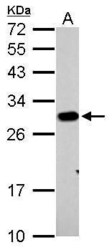
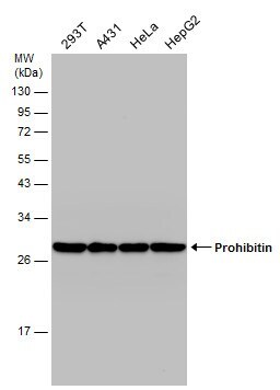
- Prohibitin Polyclonal Antibody detects PHB protein by western blot analysis. A. 30 µg PC-12 whole cell lysate/extract.12% SDS-PAGE. Prohibitin Polyclonal Antibody (Product # PA5-27329) dilution: 1:1,000. The HRP-conjugated anti-rabbit IgG antibody was used to detect the primary antibody.
- Submitted by
- Invitrogen Antibodies (provider)
- Main image
- Experimental details
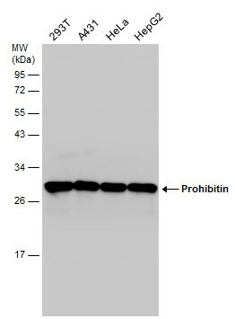
- Western Blot using Prohibitin Polyclonal Antibody (Product # PA5-27329). Various whole cell extracts (30 µg) were separated by 12% SDS-PAGE, and the membrane was blotted with Prohibitin Polyclonal Antibody (Product # PA5-27329) diluted at 1:1,000.
- Submitted by
- Invitrogen Antibodies (provider)
- Main image

- Experimental details
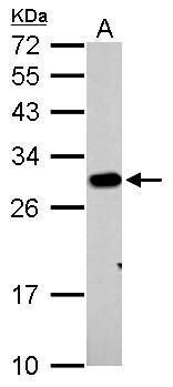
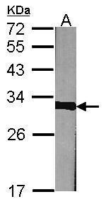
- Western Blot using Prohibitin Polyclonal Antibody (Product # PA5-27329). Sample (30 µg of whole cell lysate). Lane A: NIH-3T3. 12% SDS PAGE. Prohibitin Polyclonal Antibody (Product # PA5-27329) diluted at 1:1,000. The HRP-conjugated anti-rabbit IgG antibody was used to detect the primary antibody.
- Submitted by
- Invitrogen Antibodies (provider)
- Main image
- Experimental details
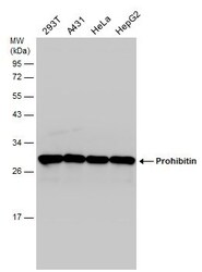
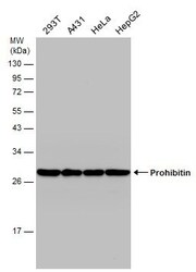
- Western Blot analysis of Prohibitin was performed by separating 30 µg of various whole cell extracts by 12% SDS-PAGE. Proteins were transferred to a membrane and probed with a Prohibitin Polyclonal Antibody (Product # PA5-27329) at a dilution of 1:1000.
Supportive validation
- Submitted by
- Invitrogen Antibodies (provider)
- Main image
- Experimental details
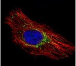
- Immunofluorescent analysis of Prohibitin in methanol-fixed HeLa cells using a Prohibitin polyclonal antibody (Product # PA5-27329) (Green) at a 1:500 dilution. Alpha-tubulin filaments were labeled with Product # PA5-29281 (Red) at a 1:2000.

- Submitted by
- Invitrogen Antibodies (provider)
- Main image
- Experimental details
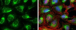
- Immunocytochemistry-Immunofluorescence analysis of Prohibitin was performed in HeLa cells fixed in 4% paraformaldehyde at RT for 15 min. Green: Prohibitin Polyclonal Antibody (Product # PA5-27329) diluted at 1:500. Red: Phalloidin, a cytoskeleton marker. Blue: Hoechst 33342 staining.

Supportive validation
- Submitted by
- Invitrogen Antibodies (provider)
- Main image

- Experimental details
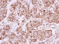
- Immunohistochemical analysis of paraffin-embedded OVCAR3 xenograft, using Prohibitin (Product # PA5-27329) antibody at 1:500 dilution. Antigen Retrieval: EDTA based buffer, pH 8.0, 15 min.

- Submitted by
- Invitrogen Antibodies (provider)
- Main image
- Experimental details
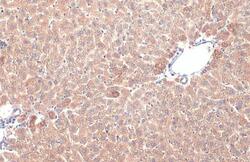
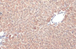
- Prohibitin Polyclonal Antibody detects Prohibitin protein at mitochondria by immunohistochemical analysis. Sample: Paraffin-embedded mouse liver. Prohibitin stained by Prohibitin Polyclonal Antibody (Product # PA5-27329) diluted at 1:500. Antigen Retrieval: Citrate buffer, pH 6.0, 15 min.

- Submitted by
- Invitrogen Antibodies (provider)
- Main image

- Experimental details
- Immunohistochemistry (Paraffin) analysis of Prohibitin was performed in paraffin-embedded human breast carcinoma tissue using Prohibitin Polyclonal Antibody (Product # PA5-27329) at a dilution of 1:500.

- Submitted by
- Invitrogen Antibodies (provider)
- Main image
- Experimental details
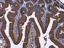
- Immunohistochemistry (Paraffin) analysis of Prohibitin was performed in paraffin-embedded mouse intestine tissue using Prohibitin Polyclonal Antibody (Product # PA5-27329) at a dilution of 1:500.

- Submitted by
- Invitrogen Antibodies (provider)
- Main image
- Experimental details
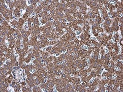
- Immunohistochemistry (Paraffin) analysis of Prohibitin was performed in paraffin-embedded rat liver tissue using Prohibitin Polyclonal Antibody (Product # PA5-27329) at a dilution of 1:500.

- Submitted by
- Invitrogen Antibodies (provider)
- Main image
- Experimental details
- Prohibitin Polyclonal Antibody detects Prohibitin protein at mitochondria by immunohistochemical analysis. Sample: Paraffin-embedded mouse liver. Prohibitin stained by Prohibitin Polyclonal Antibody (Product # PA5-27329) diluted at 1:500. Antigen Retrieval: Citrate buffer, pH 6.0, 15 min.

- Submitted by
- Invitrogen Antibodies (provider)
- Main image
- Experimental details
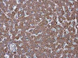
- Immunohistochemistry (Paraffin) analysis of Prohibitin was performed in paraffin-embedded human breast carcinoma tissue using Prohibitin Polyclonal Antibody (Product # PA5-27329) at a dilution of 1:500.

- Submitted by
- Invitrogen Antibodies (provider)
- Main image
- Experimental details
- Immunohistochemistry (Paraffin) analysis of Prohibitin was performed in paraffin-embedded rat liver tissue using Prohibitin Polyclonal Antibody (Product # PA5-27329) at a dilution of 1:500.

Supportive validation
- Submitted by
- Invitrogen Antibodies (provider)
- Main image
- Experimental details
- NULL

- Submitted by
- Invitrogen Antibodies (provider)
- Main image
- Experimental details
- NULL
- Submitted by
- Invitrogen Antibodies (provider)
- Main image
- Experimental details
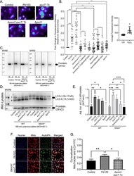
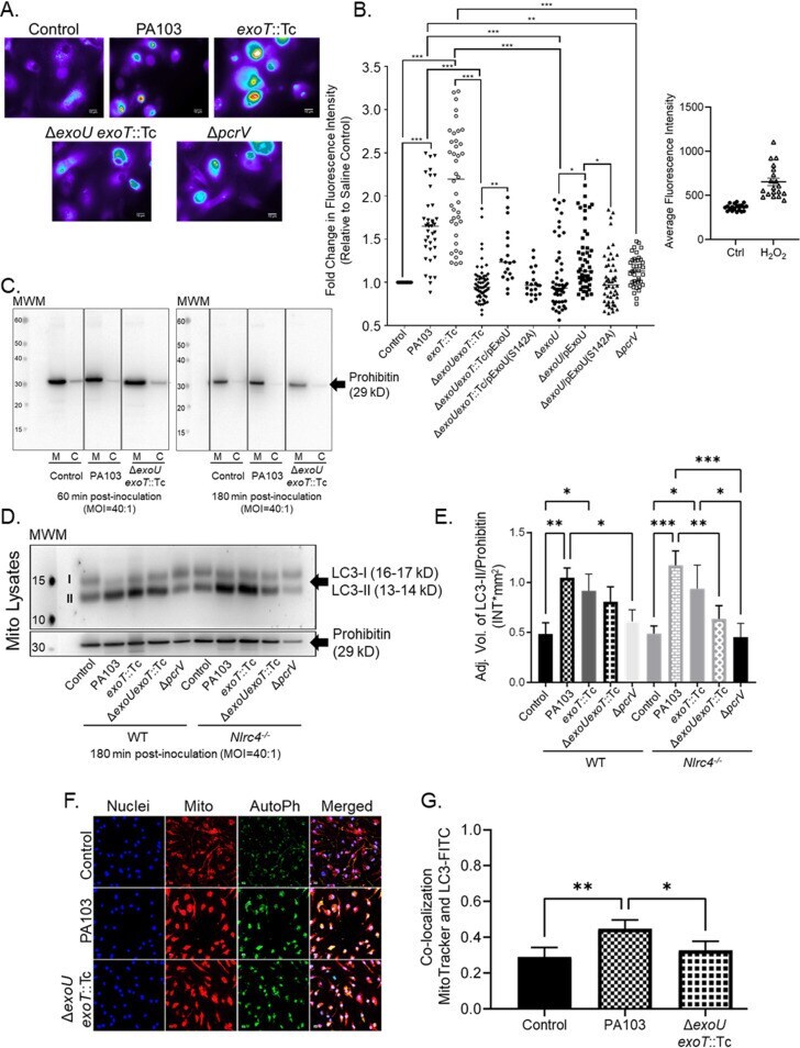
- FIG 2 ExoU elicits oxidative stress and induces autophagy at the mitochondria during P. aeruginosa infection. (A) MitoSOX-labeled WT BMDMs were treated with sterile saline (control) or inoculated with P. aeruginosa strain PA103, PA103 exoT ::Tc, PA103 Delta exoU exoT ::Tc, or PA103 Delta pcrV at an MOI of 20:1. At 180 min postinoculation, images were acquired by fluorescence microscopy. Images are representative of 3 to 7 biological replicates. The scale bar is equal to 10 mum. (B) Average fluorescence intensity of images from panel A, including additional strains as indicated. Data for several of the groups were determined to be non-normally distributed (D'Agostino and Pearson normality test); thus, all groups were compared by one-way ANOVA with Kruskal-Wallis post hoc test. All statistical comparison data are reported in Table S1. As a control, WT BMDMs were treated with either cDMEM (Ctrl) or H 2 O 2 (400 muM) for 180 min. Images were acquired by fluorescence microscopy and intensities determined (2 biological replicates). (C) WT BMDMs treated with sterile saline (control) or inoculated with P. aeruginosa strain PA103 or PA103 Delta exoU exoT ::Tc for 60 min or 180 min. Enriched mitochondrial (M) and cytosol (C) fractions (2 mug of total protein) were assayed by Western blotting to measure prohibitin as a marker of mitochondrial enrichment (29 kDa). Blots were run on separate gels and images compiled. (D) WT or Nlrc4 -/- BMDMs were treated with sterile saline (control) or
- Submitted by
- Invitrogen Antibodies (provider)
- Main image

- Experimental details
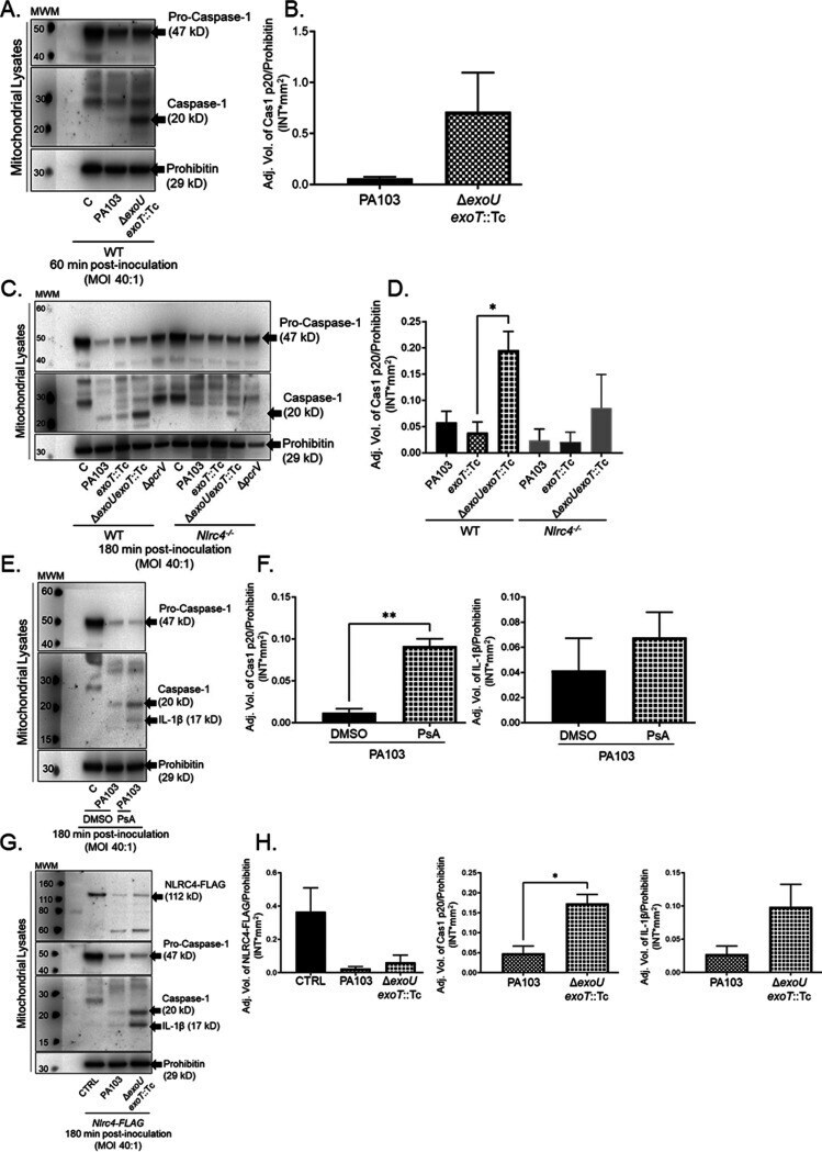
- FIG 4 ExoU inhibits caspase-1 activation at the mitochondria. (A) WT BMDMs were treated with sterile saline (C) or inoculated with P. aeruginosa strains PA103 or PA103 Delta exoU exoT ::Tc. At 60 min postinoculation, enriched mito-MAM fractions were isolated, and pro-caspase-1, active caspase-1, and prohibitin were measured by Western blotting. (B) Densitometry analysis of blots shown in panel A, where pro-caspase-1 and active caspase-1 levels were normalized to the loading control (prohibitin). (C) WT and Nlrc4 -/- BMDMs were treated with sterile saline or inoculated with P. aeruginosa strain PA103, PA103 exoT ::Tc, PA103 Delta exoU exoT ::Tc, or PA103 Delta pcrV . At 180 min postinoculation, enriched mito-MAM fractions were isolated for Western blot analysis. (D) Densitometry analysis of blots shown in panel C, where pro-caspase-1 and active caspase-1 levels were normalized to the loading control (prohibitin). * , P value = 0.03. (E) WT BMDMs were preincubated with DMSO (vehicle control) or the ExoU-specific inhibitor PsA (50 muM), where compounds were added 60 min prior to inoculation. Subsequently, cultures were treated with sterile saline or inoculated with P. aeruginosa strain PA103 for 180 min (DMSO or PsA was maintained throughout the time course). Enriched mito-MAM fractions were isolated, and pro-caspase-1, active caspase-1, active IL-1beta, and prohibitin were measured by Western blotting. (F) Densitometry analysis of caspase-1 (top) and IL-1beta (bottom) from the