Explore
 Validate
 Learn
Learn Western blot
Antibody data
- Antibody Data
- Antigen structure
- References [0]
- Comments [0]
- Validations
- Western blot [2]
- ELISA [1]
- Immunocytochemistry [1]
- Immunohistochemistry [4]
- Flow cytometry [1]
Submit
Validation data
Reference
Comment
Report error
- Product number
- AM06675SU-N - Provider product page

- Provider
- Acris Antibodies GmbH
- Proper citation
- Acris Antibodies GmbH Cat#AM06675SU-N, RRID:AB_11217208
- Product name
- anti Prohibitin / PHB
- Antibody type
- Monoclonal
- Antigen
- Purified recombinant fragment of human PHB expressed in E. Coli.
- Reactivity
- Human, Mouse, Rat, Simian
- Host
- Mouse
- Isotype
- IgG
- Antibody clone number
- 5H7
- Vial size
- 0.1 ml
No comments: Submit comment
Supportive validation
- Submitted by
- Acris Antibodies GmbH (provider)
- Main image
- Experimental details
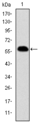
- Western blot analysis using PHB mAb against human PHB (AA: 68-259) recombinant protein. (Expected MW is 46.7 kDa)

- Submitted by
- Acris Antibodies GmbH (provider)
- Main image

- Experimental details
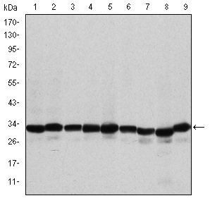
- Western blot analysis using PHB mouse mAb against A431 (1), MCF-7 (2), Jurkat (3), Hela (4), HepG2 (5), A549 (6), NIH/3T3 (7), Cos7 (8) and PC-12 (9) cell lysate.
Supportive validation
- Submitted by
- Acris Antibodies GmbH (provider)
- Main image
- Experimental details
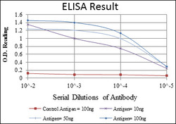
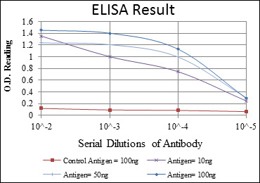
- Red: Control Antigen (100ng)Purple: Antigen (10ng)Green: Antigen (50ng)Blue: Antigen (100ng)
Supportive validation
- Submitted by
- Acris Antibodies GmbH (provider)
- Main image
- Experimental details
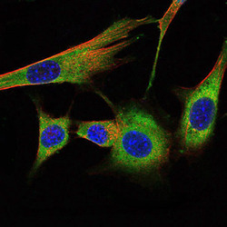
- Immunofluorescence analysis of NIH/3T3 cells using PHB mouse mAb (green). Blue: DRAQ5 fluorescent DNA dye. Red: Actin filaments have been labeled with Alexa Fluor-555 phalloidin.

Supportive validation
- Submitted by
- Acris Antibodies GmbH (provider)
- Main image
- Experimental details
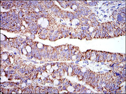
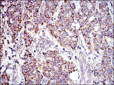
- Immunohistochemical analysis of paraffin-embedded rectum cancer tissues using PHB mouse mAb with DAB staining.

- Submitted by
- Acris Antibodies GmbH (provider)
- Main image
- Experimental details
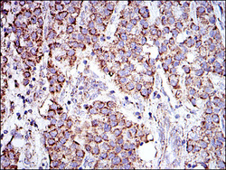
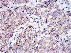
- Immunohistochemical analysis of paraffin-embedded liver cancer tissues using PHB mouse mAb with DAB staining.
- Submitted by
- Acris Antibodies GmbH (provider)
- Main image
- Experimental details
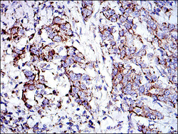
- Immunohistochemical analysis of paraffin-embedded lung cancer tissues using PHB mouse mAb with DAB staining.

- Submitted by
- Acris Antibodies GmbH (provider)
- Main image
- Experimental details
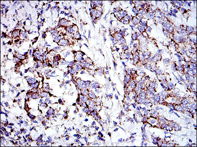
- Immunohistochemical analysis of paraffin-embedded stomach cancer tissues using PHB mouse mAb with DAB staining.
Supportive validation
- Submitted by
- Acris Antibodies GmbH (provider)
- Main image

- Experimental details
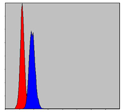
- Flow cytometric analysis of MCF-7 cells using PHB mouse mAb (blue) and negative control (red).