Explore
 Validate
 Learn
Learn Western blot
 Immunocytochemistry
Antibody data
- Antibody Data
- Antigen structure
- References [5]
- Comments [0]
- Validations
- Immunocytochemistry [1]
Submit
Validation data
Reference
Comment
Report error
- Product number
- HPA003280 - Provider product page

- Provider
- Atlas Antibodies
- Proper citation
- Atlas Antibodies Cat#HPA003280, RRID:AB_1855732
- Product name
- Anti-PHB
- Antibody type
- Polyclonal
- Description
- Polyclonal Antibody against Human PHB, Gene description: prohibitin, Alternative Gene Names: PHB1, Validated applications: WB, ICC, IHC, Uniprot ID: P35232, Storage: Store at +4°C for short term storage. Long time storage is recommended at -20°C.
- Reactivity
- Human, Mouse, Rat
- Host
- Rabbit
- Conjugate
- Unconjugated
- Isotype
- IgG
- Vial size
- 100 µl
- Concentration
- 0.1 mg/ml
- Storage
- Store at +4°C for short term storage. Long time storage is recommended at -20°C.
- Handling
- The antibody solution should be gently mixed before use.
Submitted references Fatty acid mobilization from adipose tissue is mediated by CD36 posttranslational modifications and intracellular trafficking
Prohibitin/annexin 2 interaction regulates fatty acid transport in adipose tissue
Automated Learning of Subcellular Variation among Punctate Protein Patterns and a Generative Model of Their Relation to Microtubules
The unique stem cell system of the immortal larva of the human parasite Echinococcus multilocularis
Immunofluorescence and fluorescent-protein tagging show high correlation for protein localization in mammalian cells
Daquinag A, Gao Z, Fussell C, Immaraj L, Pasqualini R, Arap W, Akimzhanov A, Febbraio M, Kolonin M
JCI Insight 2021;6(17)
JCI Insight 2021;6(17)
Prohibitin/annexin 2 interaction regulates fatty acid transport in adipose tissue
Salameh A, Daquinag A, Staquicini D, An Z, Hajjar K, Pasqualini R, Arap W, Kolonin M
JCI Insight 2016;1(10)
JCI Insight 2016;1(10)
Automated Learning of Subcellular Variation among Punctate Protein Patterns and a Generative Model of Their Relation to Microtubules
Tresch A, Johnson G, Li J, Shariff A, Rohde G, Murphy R
PLOS Computational Biology 2015;11(12):e1004614
PLOS Computational Biology 2015;11(12):e1004614
The unique stem cell system of the immortal larva of the human parasite Echinococcus multilocularis
Koziol U, Rauschendorfer T, Zanon Rodríguez L, Krohne G, Brehm K
EvoDevo 2014;5(1)
EvoDevo 2014;5(1)
Immunofluorescence and fluorescent-protein tagging show high correlation for protein localization in mammalian cells
Stadler C, Rexhepaj E, Singan V, Murphy R, Pepperkok R, Uhlén M, Simpson J, Lundberg E
Nature Methods 2013;10(4):315-323
Nature Methods 2013;10(4):315-323
No comments: Submit comment
Supportive validation
- Submitted by
- Atlas Antibodies (provider)
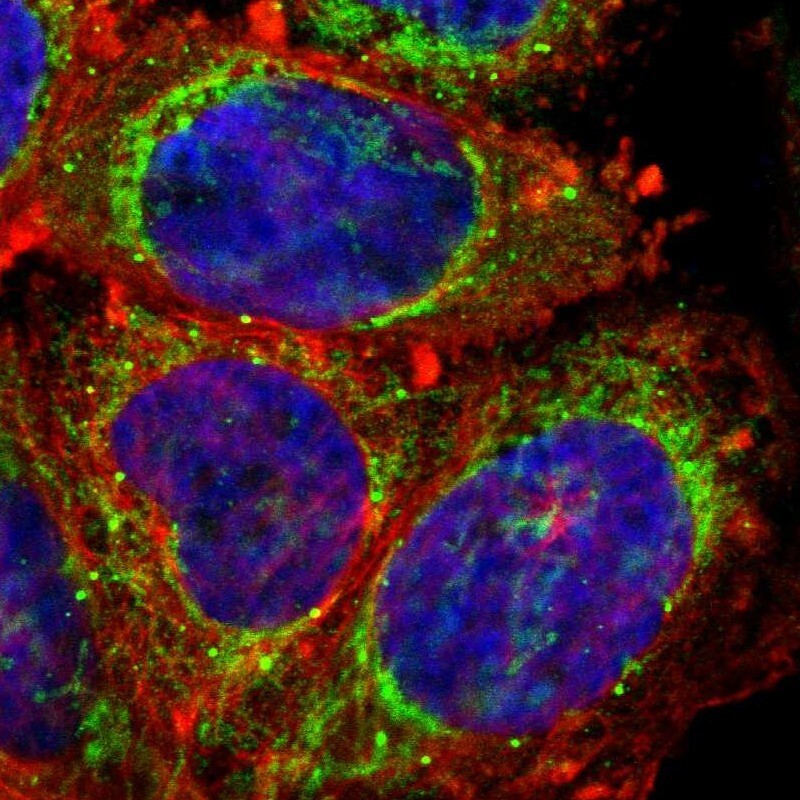
- Main image

- Experimental details
- Immunofluorescent staining of human cell line A-431 shows localization to mitochondria & vesicles.
- Sample type
- Human