Explore
 Validate
 Learn
Learn Western blot
Antibody data
- Antibody Data
- Antigen structure
- References [1]
- Comments [0]
- Validations
- Western blot [4]
- Immunoprecipitation [1]
- Immunohistochemistry [1]
Submit
Validation data
Reference
Comment
Report error
- Product number
- GTX109894 - Provider product page

- Provider
- GeneTex
- Proper citation
- GeneTex Cat#GTX109894, RRID:AB_1950821
- Product name
- Lamin B2 antibody [N3C2], Internal
- Antibody type
- Polyclonal
- Reactivity
- Human, Mouse
- Host
- Rabbit
Submitted references Long noncoding RNA LncHIFCAR/MIR31HG is a HIF-1α co-activator driving oral cancer progression.
Shih JW, Chiang WF, Wu ATH, Wu MH, Wang LY, Yu YL, Hung YW, Wang WC, Chu CY, Hung CL, Changou CA, Yen Y, Kung HJ
Nature communications 2017 Jun 22;8:15874
Nature communications 2017 Jun 22;8:15874
No comments: Submit comment
Supportive validation
- Submitted by
- GeneTex (provider)
- Main image
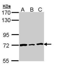
- Experimental details
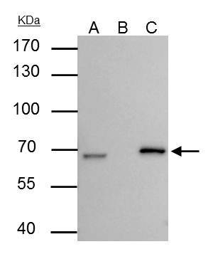
- Sample (30 ?g of whole cell lysate) A: H1299 B: HeLa C: HepG2 (GTX27900) 7.5% SDS PAGE GTX109894 diluted at 1:1000 The HRP-conjugated anti-rabbit IgG antibody (GTX213110-01) was used to detect the primary antibody.

- Submitted by
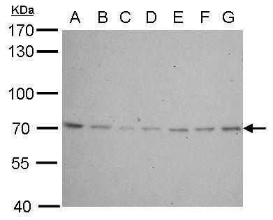
- GeneTex (provider)
- Main image

- Experimental details
- Lamin B2 antibody [N3C2], Internal detects LMNB2 protein by western blot analysis.A. 30 ?g Neuro2A whole cell lysate/extract B. 30 ?g GL261 whole cell lysate/extract C. 30 ?g C8D30 whole cell lysate/extract D. 30 ?g NIH-3T3 whole cell lysate/extract E. 30 ?g BCL-1 lysate/extract F. 30 ?g Raw264.7 whole cell lysate/extract G. 30 ?g C2C12 whole cell lysate/extract7.5% SDS-PAGELamin B2 antibody [N3C2], Internal (GTX109894) dilution: 1:500 The HRP-conjugated anti-rabbit IgG antibody (GTX213110-01) was used to detect the primary antibody.
- Submitted by
- GeneTex (provider)
- Main image
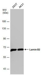
- Experimental details
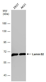
- Various whole cell extracts (30 ?g) were separated by 7.5% SDS-PAGE, and the membrane was blotted with Lamin B2 antibody [N3C2], Internal (GTX109894) diluted at 1:1000. The HRP-conjugated anti-rabbit IgG antibody (GTX213110-01) was used to detect the primary antibody.
- Submitted by
- GeneTex (provider)
- Main image
- Experimental details
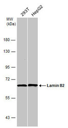
- Various whole cell extracts (30 ?g) were separated by 7.5% SDS-PAGE, and the membrane was blotted with Lamin B2 antibody [N3C2], Internal (GTX109894) diluted at 1:40000.
Supportive validation
- Submitted by
- GeneTex (provider)
- Main image

- Experimental details
- Lamin B2 antibody [N3C2], Internal immunoprecipitates Lamin B2 protein in IP experiments. IP Sample: HepG2 whole cell lysate/extract A : 30 £gg whole cell lysate/extract of Lamin B2 protein expressing HepG2 cells B : Control with 3 £gg of pre-immune rabbit IgG C : Immunoprecipitation of Lamin B2 by 3 £gg of Lamin B2 antibody [N3C2], Internal (GTX109894) 7.5% SDS-PAGE The immunoprecipitated Lamin B2 protein was detected by Lamin B2 antibody [N3C2], Internal (GTX109894) diluted at 1 : 1000. EasyBlot anti-rabbit IgG (HRP) (GTX221666-01) was used as a secondary reagent.
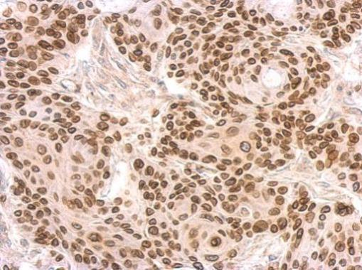
Supportive validation
- Submitted by
- GeneTex (provider)
- Main image
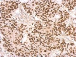
- Experimental details
- Lamin B2 antibody [N3C2], Internal detects LMNB2 protein at on Cal27 xenograft by immunohistochemical analysis. Sample: Paraffin-embedded Cal27 xenograft. Lamin B2 antibody [N3C2], Internal (GTX109894) dilution: 1:500.